

Three-dimensional rotational spinal angiography in the evaluation and treatment of vascular malformations
- PMID: 12917141
- PMCID: PMC7973676
Three-dimensional rotational spinal angiography in the evaluation and treatment of vascular malformations
Abstract
Background and purpose: Conventional spinal angiography, although useful in providing angioarchitectural details of spinal vascular disease, has limitations. The advent of 3D angiography has provided a better comprehension of angioarchitectural detail when evaluating the intracranial circulation. The purpose of this study was to evaluate the usefulness of 3D angiography in the diagnosis and treatment of vascular malformations of the spine.
Methods: This retrospective analysis included 17 3D spinal angiograms acquired in 14 consecutive patients examined at our institution for a spinal vascular lesion, which included nine spinal cord arteriovenous malformations (AVMs), one perimedullary arteriovenous fistula (AVF), three spinal dural AVFs, and one nerve root AVM. 3D angiography was obtained with apnea under general anesthesia by using a 14-second acquisition and 200 degrees rotation of the gantry during injection of 300 mg I/mL nonionic contrast material at a rate of 0.5-3.5 mL/s. Multiple reconstructed images were obtained with or without opacification of the surrounding structures. These images were then evaluated by the interventionalists at the time of the procedure and compared with findings obtained by conventional subtraction angiography.
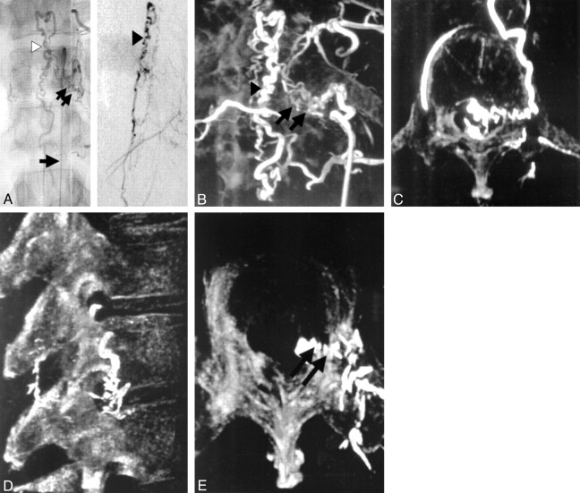
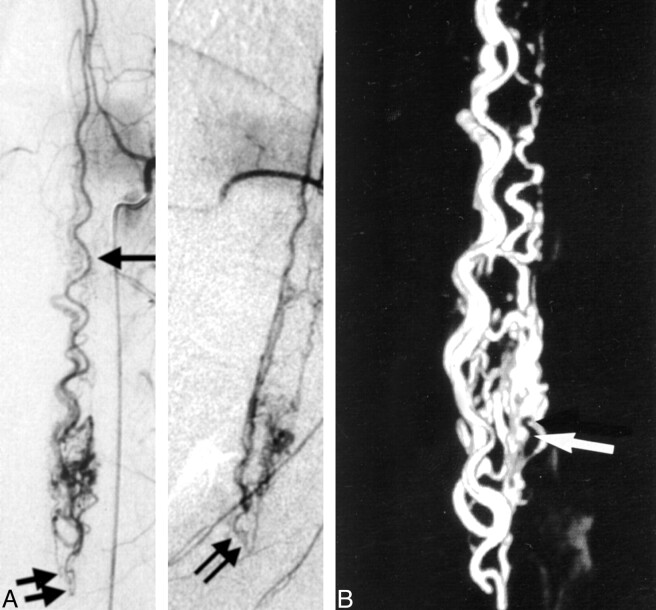
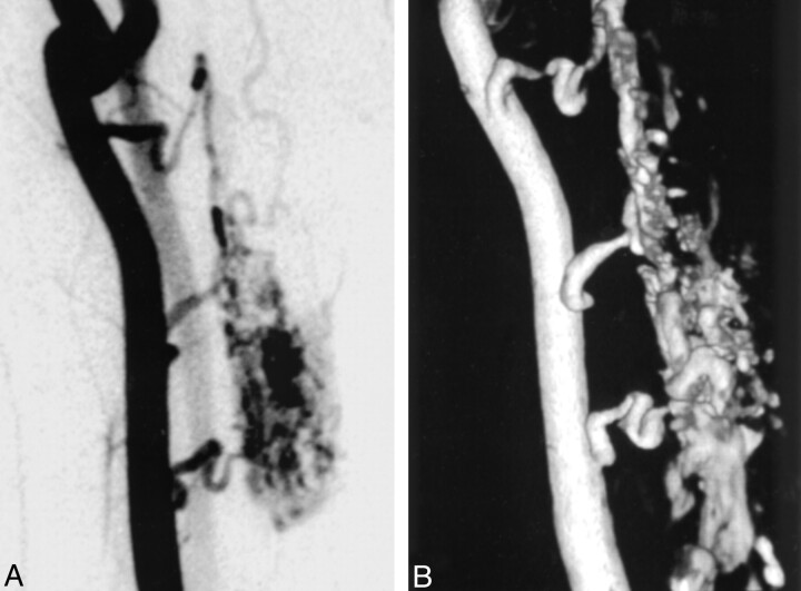
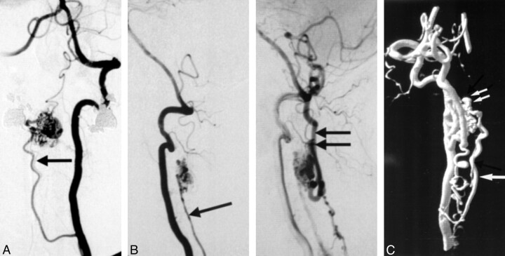
Results: 3D angiography was useful in differentiating intramedullary lesions from perimedullary surface lesions; detecting arterial, nidal, or venous aneurysms; and evaluating the 3D structure of the lesion as well as the relationship between the malformation and its draining veins or surrounding bony structures. In specific situations, it obviated the need for contrast-enhanced conventional or 3D CT, as well as for lateral or oblique angiographic views, which are sometimes difficult to obtain with good quality. No 3D angiography-related complications were experienced. Some limitations in the definition of small vessel anatomy in the reconstructed images were noted.
Conclusion: In this small series of patients, 3D angiography was safe and useful for evaluation of the 3D vascular anatomy of spinal vascular malformations.
Figures
References
-
- Gaupp J. Hamorrhoiden der Pia mater spinalis im Geibiet des Lendenmarks. Beitr Pathol 1888;2:516–518
-
- Djindjian R. Angiographie de la moelle epiniere. Acta Radiol (Diagn) 1969;9:707–726 - PubMed
-
- DiChiro G, Doppman JL, Ommaya A. Selective arteriography of arteriovenous aneurysms of the spinal cord. Radiology 1967;88:1065–1077 - PubMed
-
- Doppman JL. Arteriography of the spinal cord. Semin Roentgenol 1972;7:231–239 - PubMed
-
- Turjman F, Bendib K, Girerd C, et al. Pretherapeutic evaluation of intracranial aneurysms using three-dimensional angiography (3D morphometer): preliminary results. In: Taki W, Picard L, Kikuchi H, eds. Advances in Interventional Neuroradiology and Intravascular Neurosurgery. Amsterdam: Elsevier Science;1996;75–79
Publication types
MeSH terms
LinkOut - more resources
Full Text Sources
Medical