

Case Reports
Neuroimaging features of epidermal nevus syndrome
Affiliations
- PMID: 12917149
- PMCID: PMC7973704
Item in Clipboard
Case Reports
Neuroimaging features of epidermal nevus syndrome
AJNR Am J Neuroradiol.
2003 Aug.
Abstract
Epidermal nevus syndrome is a kind of neurocutaneous syndrome that is associated with epidermal nevus and a variety of congenital CNS disorders. Clinical presentations include seizures, paresis, mental retardation, and developmental delay. We report three cases with MR imaging and magnetoencephalography findings; one patient underwent ictal and interictal single photon emission CT. Both structural and functional imaging studies indicated that the frontal lobes had lesser involvement or were intact. One patient underwent hemispherectomy because of the medically intractable seizure. He remained seizure free with topiramate monotherapy.
Figures
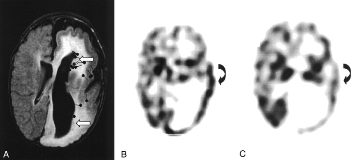
Images from the case of patient 1, a 13-month-old male patient who presented with left face epidermal nevus and right hemiparesis complicated with complex partial seizures. A, FLAIR image shows left hemimegalencephaly associated with ipsilateral enlarged ventricle, dysmyelination, and thickened and flattened cortex. Open left insula, blurred white-gray matter junction, and heterotopias (arrows) can be seen. Sources of interictal spikes (black spots) are scattered over central parietal region and deep around the heterotopia region of the left hemisphere. B, Ictal SPECT scan shows increased perfusion in the left parietal (curved arrow) and temporal lobes. C, Interictal SPECT scan of corresponding area shows decreased activity (curved arrow).

Images from the case of patient 2, a 26-month-old female patient who presented with left facial epidermal nevus and facial and neck hypertrophy. A, Photograph shows left facial hypertrophy and epidermal nevus (arrow). B, FLAIR image shows the left hemimegalencephaly, enlarged lateral ventricle, dysmyelinated white matter, blurred white-gray matter junction, and plate-like thickened cortex in the temporal, parietal, and occipital lobes. The left frontal lobe and right hemisphere are normal. Sources of interictal spikes (black spots) are scattered around the posterior part of the left hemisphere.

Axial view T2-weighted image of patient 3 shows left hemimegalencephaly sparing the frontal lobe, with appearances similar to those on the images of patient 2. Interictal epileptic sharp waves or spikes (black spots) are localized in temporal and parietal areas.
References
-
- Grebe TA, Rimsza ME, Richter SF, Hansen RC, Hoyme H. Further delineation of the epidermal nevus syndrome: two cases with new findings and literature review. Am J Med Genet 1993;47:24–30 - PubMed
-
- Pavone L, Curatolo P, Rizzo R, et al. Epidermal nevus syndrome: a neurologic variant with hemimegalencephaly, gyral malformation, mental retardation, seizures, and facial hemihypertrophy. Neurology 1991;41:266–271 - PubMed
-
- Herman TE, Siegel MJ. Hemimegalencephaly and linear nevus sebaceous syndrome. J Perinatol 2001;21:336–338 - PubMed
-
- Olivares JL, Ramos FJ, Carapeto FJ, Bueno M. Epidermal naevus syndrome and hypophosphataemic rickets: description of a patient with central nervous system anomalies and review of the literature. Eur J Pediatr 1999;158:103–107 - PubMed
-
- Hennekam RC, Kwa VI, van Amerongen A. Arteriovenous and lymphatic malformations, linear verrucous epidermal nevus and mild overgrowth: another hamartoneoplastic syndrome? Clin Dysmorphol 1999;8:111–115 - PubMed
Publication types
MeSH terms
Grants and funding
LinkOut - more resources
Full Text Sources