. 2003 Sep;23(17):6075-85.
doi: 10.1128/MCB.23.17.6075-6085.2003.

Of mice and MEN1: Insulinomas in a conditional mouse knockout

Affiliations

Of mice and MEN1: Insulinomas in a conditional mouse knockout

Judy S Crabtree et al. Mol Cell Biol. 2003 Sep.

Abstract

Patients with multiple endocrine neoplasia type 1 (MEN1) develop multiple endocrine tumors, primarily affecting the parathyroid, pituitary, and endocrine pancreas, due to the inactivation of the MEN1 gene. A conditional mouse model was developed to evaluate the loss of the mouse homolog, Men1, in the pancreatic beta cell. Men1 in these mice contains exons 3 to 8 flanked by loxP sites, such that, when the mice are crossed to transgenic mice expressing cre from the rat insulin promoter (RIP-cre), exons 3 to 8 are deleted in beta cells. By 60 weeks of age, >80% of mice homozygous for the floxed Men1 gene and expressing RIP-cre develop multiple pancreatic islet adenomas. The formation of adenomas results in elevated serum insulin levels and decreased blood glucose levels. The delay in tumor appearance, even with early loss of both copies of Men1, implies that additional somatic events are required for adenoma formation in beta cells. Comparative genomic hybridization of beta cell tumor DNA from these mice reveals duplication of chromosome 11, potentially revealing regions of interest with respect to tumorigenesis.


Figures
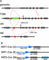
FIG. 1.
FIG. 1.
Genomic structure of Men1 alleles and RIP-cre transgenes. (A) In vivo manipulation of the Men1 gene. The genomic structure of the Men1 gene (top line) was altered by the insertion of a floxed 3-phosphoglycerate kinase (PGK)-neomycin cassette in intron 2 and a third loxP in intron 8 to generate the TSM allele in Men1TSM/+ mice, as previously reported (6). Men1TSM/+ mice were then bred to EIIa-cre transgenic mice, and the progeny were selected for loss of the PGK-neomycin cassette; the resulting allele was termed ΔN. Breeding Men1ΔN/ΔN mice to RIP-cre transgenic mice resulted in progeny with exons 3 to 8 excised in a tissue-specific manner to generate the del allele. Grey boxes, exons; red triangles, loxP sites; green box, PGK-neomycin cassette in the transcriptional orientation opposite to that for Men1; small black bars, genotyping primer locations F and G. (B) RIP-cre transgenes. Three different lines of cre-expressing mice were utilized, each with the cre recombinase under the control of a portion of the rat insulin promoter (RIP).
FIG. 2.
FIG. 2.
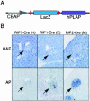
Expression pattern of Cre in pancreata of RIP-cre transgenic mice bred to Z/AP reporter mice. (A) Structure of the Z/AP reporter transgene. Z/AP reporter mice contain the chicken beta actin promoter (CBAP) followed by a floxed lacZ gene and then a heat-stable human placental AP (hPLAP) gene (23). Red triangles, loxP sites. (B) All three lines of RIP-cre mice were bred to transgenic Z/AP mice. The pancreata of 6-week-old mice carrying both transgenes were serially sectioned, and neighboring sections were stained with either hematoxylin and eosin (H&E) or AP. Arrows, pancreatic islets.
FIG. 3.
FIG. 3.
Histology and tumor incidence. (A) Pancreatic islet morphology of 24-week-old Men1ΔN/ΔN mice with one of three RIP-cre transgenes. Sections were stained with hematoxylin and eosin and photographed at 100× magnification. (B) Tumor incidence in Men1ΔN/ΔN; RIP-cre mice. Mice carrying the RIP2-cre (M) transgene develop tumors at an earlier age than either the RIP7-cre (H) or the RIP1-cre (E) mice, as evaluated by determining the percentage of mice with no tumor formation at the scheduled date of autopsy. (C) Tumor incidence in Men1ΔN/+; RIP-cre mice. Heterozygote floxed mice with a RIP-cre transgene develop tumors more slowly than the homozygotes. The rate is apparently independent of cre expression levels, as shown by the percentage of mice tumor free at the scheduled date of autopsy.
FIG.4.
FIG.4.
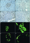
Loss of menin in beta cells. (A to D) Menin IHC with the SQV-R4 antibody. Brown nuclear staining indicates the menin protein. (A) Menin IHC on the wild-type pancreas of a 56-week-old animal. (B) Pancreas of a 60-week-old Men1ΔN/+; RIP2-cre mouse showing a possible reduction (50%) of menin in the nuclei of a hyperplastic islet, but not complete loss. (C and D) Pancreas of a 48-week-old Men1ΔN/ΔN; RIP2-cre mouse showing absence of menin in most of the nuclei of atypical islets (C) and tumors (D), while acinar tissue retains menin staining. (E to H) Dual immunofluorescence in beta cells. (E) Wild-type pancreatic islet stained for menin (red) and insulin (green). (F) Pancreas from a 4-week-old Men1ΔN/ΔN; RIP2-cre mouse showing loss of menin in insulin-positive cells and presence of menin in cells negative for insulin (arrow). (G) Wild-type pancreas stained for menin (red) and glucagon (green). (H) Pancreas from a 4-week-old Men1ΔN/ΔN; RIP2-cre mouse showing retention of menin in the cells positive for glucagon but its absence in the majority of the other islet cells.
FIG. 5.
FIG. 5.
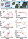
Insulin IHC and blood chemistry. (A) Insulin IHC on sections from Men1ΔN/ΔN; RIP7-cre/+ (H), Men1ΔN/ΔN; RIP1-cre/+ (E), and Men1ΔN/ΔN; RIP2-cre/+ (M) mice showing insulin production by atypical islets. (B and C) Serum insulin and blood glucose of fasted Men1 floxed mice with the indicated cre transgene. Red squares, Men1ΔN/ΔN; RIP-cre mice; green triangles, Men1ΔN/+; RIP-cre mice; gold diamonds, mice containing a cre transgene but wild-type for Men1. Error bars, standard error of the mean; asterisk, P < 0.005 by analysis of variance.
FIG. 6.
FIG. 6.
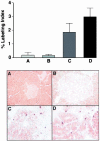
Islet proliferation as shown by BrdU labeling. Islets that appear normal in both wild-type (A) and RIP2-cre-positive (B) control mice show minimal labeling, whereas atypical islets (C) and adenomas (D) show increased BrdU labeling. This suggests that increased cellular proliferation contributes to tumorigenesis.
FIG. 7.
FIG. 7.
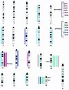
CGH of menin null tumors. CGH was performed on tumor samples from four Men1ΔN/ΔN; RIP2-cre/+ mice (M1 to M4) and seven conventional heterozygote knockout mice (KO1 to KO7). Pancreatic samples show gain of chromosome 11, whereas pituitary samples show duplication of chromosome 15, regardless of genotype. Conventional knockout samples have the expected loss of chromosome 19 and, thus, LOH for the Men1 locus. Bars to the left of chromosomes indicate chromosomal loss, and bars to the right indicate chromosomal gains.
FIG. 8.
FIG. 8.
Model of Men1 function in islets. Stage 1 shows wild-type islets (10×). Stage 2 is a uniform population of hyperplastic islet cells (20×). Stage 3 is atypical islet hyperplasia, showing pleomorphism of hyperplastic islet cells (20×), and stage 4 shows the pseudorosette pattern typical of adenoma. Loss of one Men1 allele, such as in the conventional heterozygote knockout or the heterozygote conditional mice with RIP-cre, leads to islet hyperplasia over time. Loss of the second allele, either by homozygous conditional targeting in the presence of RIP-cre or by LOH (for conventional heterozygote knockout of Men1), leads to atypia. Subsequent somatic events are required for tumor formation.

References

    1. Agarwal, S. K., L. V. Debelenko, M. B. Kester, S. C. Guru, P. Manickam, S. E. Olufemi, M. C. Skarulis, C. Heppner, J. S. Crabtree, I. A. Lubensky, Z. Zhuang, Y. S. Kim, S. C. Chandrasekharappa, F. S. Collins, L. A. Liotta, A. M. Spiegel, A. L. Burns, M. R. Emmert-Buck, and S. J. Marx. 1998. Analysis of recurrent germline mutations in the MEN1 gene encountered in apparently unrelated families. Hum. Mutat. 12:75-82. - PubMed
    1. Agarwal, S. K., S. C. Guru, C. Heppner, M. R. Erdos, R. M. Collins, S. Y. Park, S. Saggar, S. C. Chandrasekharappa, F. S. Collins, A. M. Spiegel, S. J. Marx, and A. L. Burns. 1999. Menin interacts with the AP1 transcription factor JunD and represses JunD-activated transcription. Cell 96:143-152. - PubMed
    1. Ahlgren, U., J. Jonsson, L. Jonsson, K. Simu, and H. Edlund. 1998. Beta-cell-specific inactivation of the mouse Ipf1/Pdx1 gene results in loss of the beta-cell phenotype and maturity onset diabetes. Genes Dev. 12:1763-1768. - PMC - PubMed
    1. Bassett, J. H., S. A. Forbes, A. A. Pannett, S. E. Lloyd, P. T. Christie, C. Wooding, B. Harding, G. M. Besser, C. R. Edwards, J. P. Monson, J. Sampson, J. A. Wass, M. H. Wheeler, and R. V. Thakker. 1998. Characterization of mutations in patients with multiple endocrine neoplasia type 1. Am. J. Hum Genet. 62:232-244. - PMC - PubMed
    1. Chandrasekharappa, S. C., S. C. Guru, P. Manickam, S. E. Olufemi, F. S. Collins, M. R. Emmert-Buck, L. V. Debelenko, Z. Zhuang, I. A. Lubensky, L. A. Liotta, J. S. Crabtree, Y. Wang, B. A. Roe, J. Weisemann, M. S. Boguski, S. K. Agarwal, M. B. Kester, Y. S. Kim, C. Heppner, Q. Dong, A. M. Spiegel, A. L. Burns, and S. J. Marx. 1997. Positional cloning of the gene for multiple endocrine neoplasia-type 1. Science 276:404-407. - PubMed

Publication types

MeSH terms