. 2003 Sep;23(17):6200-9.
doi: 10.1128/MCB.23.17.6200-6209.2003.

Potentiation of tumor necrosis factor-induced NF-kappa B activation by deacetylase inhibitors is associated with a delayed cytoplasmic reappearance of I kappa B alpha

Affiliations

Potentiation of tumor necrosis factor-induced NF-kappa B activation by deacetylase inhibitors is associated with a delayed cytoplasmic reappearance of I kappa B alpha

Emmanuelle Adam et al. Mol Cell Biol. 2003 Sep.

Erratum in

  • Mol Cell Biol. 2004 Aug;24(15):6890

Abstract

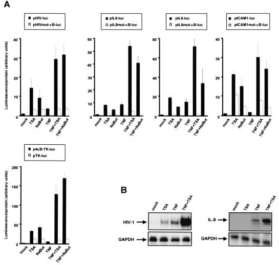
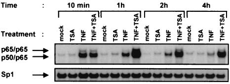
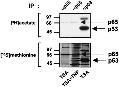
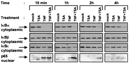
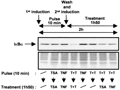
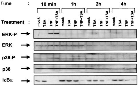
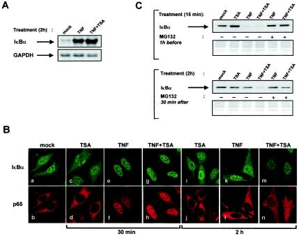
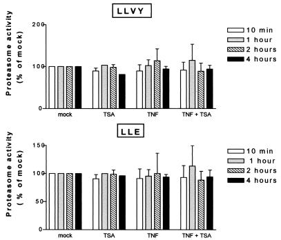
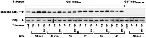
Previous studies have implicated acetylases and deacetylases in regulating the transcriptional activity of NF-kappa B. Here, we show that inhibitors of deacetylases such as trichostatin A (TSA) and sodium butyrate (NaBut) potentiated TNF-induced expression of several natural NF-kappa B-driven promoters. This transcriptional synergism observed between TNF and TSA (or NaBut) required intact kappa B sites in all promoters tested and was biologically relevant as demonstrated by RNase protection on two instances of endogenous NF-kappa B-regulated gene transcription. Importantly, TSA prolonged both TNF-induced DNA-binding activity and the presence of NF-kappa B in the nucleus. We showed that the p65 subunit of NF-kappa B was acetylated in vivo. However, this acetylation was weak, suggesting that other mechanisms could be implicated in the potentiated binding and transactivation activities of NF-kappa B after TNF plus TSA versus TNF treatment. Western blot and immunofluorescence confocal microscopy experiments revealed a delay in the cytoplasmic reappearance of the I kappa B alpha inhibitor that correlated temporally with the prolonged intranuclear binding and presence of NF-kappa B. This delay was due neither to a defect in I kappa B alpha mRNA production nor to a nuclear retention of I kappa B alpha but was rather due to a persistent proteasome-mediated degradation of I kappa B alpha. A prolongation of I kappa B kinase activity could explain, at least partially, the delayed I kappa B alpha cytoplasmic reappearance observed in presence of TNF plus TSA.


Figures

FIG. 1.
FIG. 1.
Deacetylase inhibitors potentiate TNF-induced NF-κB-driven gene expression. (A) HeLa cells were transiently transfected with 500 ng of the indicated luciferase reporter constructs. Cells were treated with TSA, NaBut, TNF, TNF plus TSA, or TNF plus NaBut (or were untreated), and cellular lysates were tested for luciferase activity. The mock-treated value of each wild-type reporter construct was arbitrarily set to a value of 1. Values represent the means of triplicate samples. A representative experiment of three independent transfections is shown. (B) RPA after a 6-h treatment of the latently HIV-1-infected cell line U1 with TNF and/or TSA. To detect HIV-1 RNA, total RNA samples were incubated with an antisense riboprobe corresponding to the HIV-1 LTR (left panel). RPA after a 2-h treatment of HeLa cells with TNF and/or TSA. Total cellular RNA was harvested after the indicated treatments and used in an RPA with a human IL-8 gene-specific riboprobe (right panel). GAPDH is shown as loading controls.
FIG. 2.
FIG. 2.
EMSA analysis of NF-κB binding activity after TNF plus TSA versus TNF treatment. Nuclear extracts were prepared from HeLa cells treated with TNF and/or TSA (or untreated) for various times. An oligonucleotide corresponding to the HIV-1-κB sites was used as the probe. As control for equal loading, the lower panel shows comparability of the various nuclear extracts assessed by EMSA with an Sp1 consensus probe.
FIG. 3.
FIG. 3.
p65 is not significantly acetylated in vivo. COS-7 cells were transfected with a p65- or p53-expression vector and labeled either with [3H]sodium acetate or [35S]methionine. Whole-cell extracts from transfected cells were immunoprecipitated with an anti-p65 or anti-p53 antibody. The immunoprecipitated proteins were analyzed by SDS-PAGE followed by autoradiography.
FIG. 4.
FIG. 4.
Delay in the cytoplasmic IκBα reappearance in response to TNF plus TSA versus TNF treatment. Cytoplasmic and nuclear extracts were prepared from HeLa cells treated with TNF and/or TSA (or untreated) for various times. Equal amounts of proteins were analyzed by Western blotting with antibodies against IκBα, IκBβ, IκBɛ, and p65.
FIG. 5.
FIG. 5.
The continuous presence of both TNF and TSA is required for the delayed cytoplasmic reappearance of IκBα. HeLa cells were pulsed for 10 min with TSA, TNF, or a combination of both. Medium was then removed after this initial 10-min treatment (i.e., after IκBα disappearance), and the cells were washed twice with PBS and then replenished for the remainder of the 2-h period (i.e., for 1 h 50 min) either with fresh medium or with fresh medium supplemented with TSA, TNF, or both. Cytoplasmic extracts were then prepared and equal amounts of proteins were resolved on 10% SDS-PAGE and immunoblotted with an anti-IκBα antibody. Coomassie blue-stained membrane after immunoblotting (bottom panel) is shown as a loading control.
FIG. 6.
FIG. 6.
TSA does not affect the activation by TNF of the p38 and ERK MAP kinases. Whole-cell extracts were prepared from HeLa cells treated with TNF and/or TSA (or untreated) for various times. Equal amounts of proteins were analyzed by Western blotting with the anti-phospho-p38 or anti-phospho-ERK antibody. Blots were reprobed with anti-p38 or anti-ERK to verify equal loading.
FIG. 7.
FIG. 7.
The delayed cytoplasmic reappearance of IκBα appears to result from a persistent proteasome-mediated degradation of IκBα. (A) The delayed cytoplasmic reappearance of IκBα is not due to a defect in IκBα mRNA production. Total RNA was isolated from HeLa cells treated with TNF or TNF plus TSA for 2 h (or untreated), and IκBα mRNA was analyzed by RPA with a human IκBα gene-specific riboprobe. GAPDH is shown as loading controls. (B) The delayed cytoplasmic reappearance of IκBα is not due to a nuclear retention of IκBα. HeLa cells were treated with TNF and/or TSA (or untreated) and analyzed by confocal microscopy at the indicated time points. Endogenous IκBα and p65 were localized by indirect immunofluorescence. (C) The delayed cytoplasmic reappearance of IκBα appears to result from a persistent proteasome-mediated degradation of IκBα. Cytoplasmic extracts were prepared from HeLa cells treated for 15 min with TNF and/or TSA as indicated after a 1-h pretreatment with MG132 (25 μM) and then analyzed for the presence of IκBα by Western blotting (top). Cytoplasmic extracts were prepared from HeLa cells treated for 2 h with TNF and/or TSA as indicated. After 30 min of this initial treatment, MG132 (25 μM) was added for the rest of the 2-h period. Extracts were immunoblotted with anti-IκBα (bottom). The Coomassie blue-stained membrane after immunoblotting is shown as a loading control.
FIG. 8.
FIG. 8.
TSA does not affect proteasome activity in presence or absence of TNF. Cell lysates were prepared from HeLa cells treated with TNF and/or TSA (or untreated) for various times and analyzed for proteasome activity. Chymotrypsin-like and peptidyl glutamyl peptide hydrolase proteasome activities were measured in lysates by using the fluorogenic peptides LLVY-AMC and LLE-NA, respectively. The proteasome inhibitor MG132 (200 μM) was used to ensure that measured activities were due to the proteasome. The mock-treated value at each time point was arbitrarily set to a value of 100%. Values represent the means of triplicate samples.
FIG. 9.
FIG. 9.
TSA prolongs TNF-induced IKK kinase activity. Endogenous IKK activity was measured in HeLa cells treated with TNF and/or TSA for different periods of time. IKKs were immunoprecipitated with an anti-IKKγ antibody, and associated kinase activity was assayed by using purified GST-IκBα1-54 fusion protein as a substrate (top). The presence of equal amounts of the IKK catalytic subunit, IKKβ, was confirmed in each sample by Western blotting (bottom). Equivalent GST-IκBα loading was verified by staining the membrane with Coomassie brilliant blue after immunoblotting (data not shown).

Similar articles

Cited by

References

    1. Aoudjit, F., N. Brochu, B. Belanger, C. Stratowa, J. Hiscott, and M. Audette. 1997. Heterodimeric retinoic acid receptor-β and retinoid X receptor-α complexes stimulate expression of the intercellular adhesion molecule-1 gene. Cell Growth Differ. 8:335-342. - PubMed
    1. Arenzana-Seisdedos, F., P. Turpin, M. Rodriguez, D. Thomas, R. T. Hay, J. L. Virelizier, and C. Dargemont. 1997. Nuclear localization of IκBα promotes active transport of NF-κB from the nucleus to the cytoplasm. J. Cell Sci. 110:369-378. - PubMed
    1. Ashburner, B. P., S. D. Westerheide, and A. S. Baldwin, Jr. 2001. The p65 (RelA) subunit of NF-κB interacts with the histone deacetylase (HDAC) corepressors HDAC1 and HDAC2 to negatively regulate gene expression. Mol. Cell. Biol. 21:7065-7077. - PMC - PubMed
    1. Baldwin, A. S., Jr. 2001. Series introduction: the transcription factor NF-κB and human disease. J. Clin. Investig. 107:3-6. - PMC - PubMed
    1. Ben Neriah, Y. 2002. Regulatory functions of ubiquitination in the immune system. Nat. Immunol. 3:20-26. - PubMed

Publication types

MeSH terms

Substances