. 2003 Sep;23(17):6291-9.
doi: 10.1128/MCB.23.17.6291-6299.2003.

Vav1 dephosphorylation by the tyrosine phosphatase SHP-1 as a mechanism for inhibition of cellular cytotoxicity

Affiliations

Vav1 dephosphorylation by the tyrosine phosphatase SHP-1 as a mechanism for inhibition of cellular cytotoxicity

Christopher C Stebbins et al. Mol Cell Biol. 2003 Sep.

Abstract

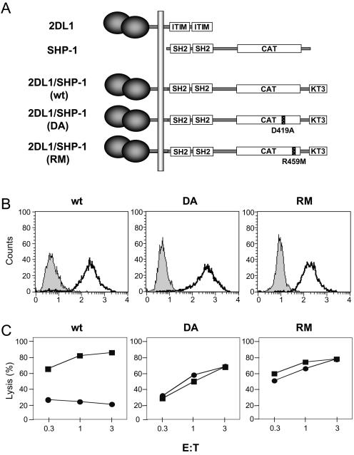
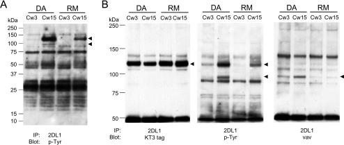
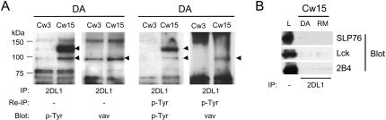
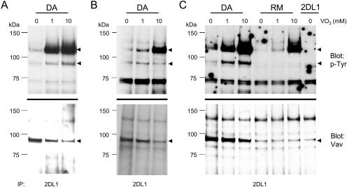
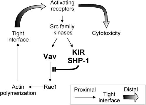
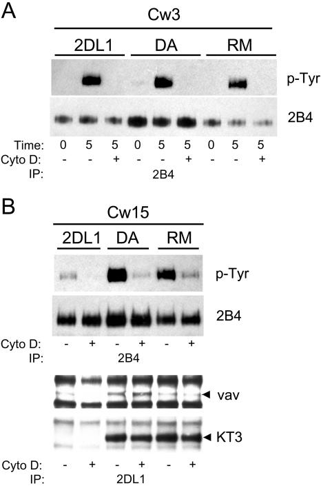
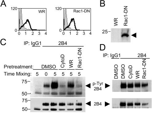
Here, we present data suggesting a novel mechanism for regulation of natural killer (NK) cell cytotoxicity through inhibitory receptors. Interaction of activation receptors with their ligands on target cells induces cytotoxicity by NK cells. This activation is under negative control by inhibitory receptors that recruit tyrosine phosphatase SHP-1 upon binding major histocompatibility class I on target cells. How SHP-1 blocks the activation pathway is not known. To identify SHP-1 substrates, an HLA-C-specific inhibitory receptor fused to a substrate-trapping mutant of SHP-1 was expressed in NK cells. Phosphorylated Vav1, a regulator of actin cytoskeleton, was the only protein detectably associated with the catalytic site of SHP-1 during NK cell contact with target cells expressing HLA-C. Vav1 trapping was independent of actin polymerization, suggesting that inhibition of cellular cytotoxicity occurs through an early dephosphorylation of Vav1 by SHP-1, which blocks actin-dependent activation signals. Such a mechanism explains how inhibitory receptors can block activating signals induced by different receptors.


Figures

FIG. 1.
FIG. 1.
Expression and function of chimeric 2DL1/SHP-1 molecules. (A) Chimeric 2DL1/SHP-1 molecules. Wild-type KIR2DL1 (2DL1) and SHP-1 are shown with the SH2 domains of SHP-1 aligned with the cytoplasmic ITIMs of 2DL1. The catalytic domain (CAT) of SHP-1 and the KT3 tag are boxed. (B) Staining of YTS cells expressing these chimeric constructs. Shaded profiles represent fluorescein isothiocyanate-labeled goat anti-mouse Ab only. Dark lines represent staining with the anti-2DL1 mAb EB6. (C) Lysis of 221-Cw3 (▪) or 221-Cw4 (•) by YTS-2DL1/SHP-1(wt), YTS-2DL1/SHP-1(RM), and YTS-2DL1/SHP-1(DA) cells at the effector-to-target ratios (E:T) indicated.
FIG. 2.
FIG. 2.
Specific association of molecules with 2DL1/SHP-1(DA) after incubation with 221-Cw15 cells. 2DL1/SHP1(DA) and 2DL1/SHP1(RM) cells were mixed for 5 min with either 221-Cw3 or 221-Cw15 cells as indicated at the top. (A) Anti-2DL1 immunoprecipitates were separated by 4 to 20% SDS-PAGE, transferred to a polyvinylidene difluoride (PVDF) membrane and Western blotted with antiphosphotyrosine mAb 4G10. Arrowheads point to two bands that are selectively enhanced in the DA-Cw15 combination. (B) Anti-2DL1 immunoprecipitates separated by 6% SDS-PAGE, transferred to a PVDF membrane, and subjected to Western blotting with antiphosphotyrosine mAb 4G10 (middle panel). After being stripped, the blot was reprobed with either an anti-Vav1 mAb (right panel) or an anti-KT3-tag mAb (left panel). Arrowheads point to the positions of tyrosine-phosphorylated proteins.
FIG. 3.
FIG. 3.
Vav1 is the only detectable tyrosine-phosphorylated protein associated with 2DL1/SHP-1(DA) after incubation with 221-Cw15 cells. 2DL1/SHP-1(DA) and 2DL1/SHP-1(RM) cells were mixed for 5 min with 221-Cw3 or 221-Cw15 cells as indicated at the top. (A) One-fourth of the immunoprecipitations with EB6 was analyzed exactly as described in the legend of Fig. 2. The remaining three-fourths was boiled in 2% SDS, diluted with lysis buffer to 0.1% SDS, reimmunoprecipitated with 4G10-agarose, and separated on a 4 to 20% SDS-polyacrylamide gel. After transfer to PVDF membranes, Western blotting was performed sequentially with anti-p-Tyr mAb 4G10 and anti-Vav1 mAb (right panels). (B) 2DL1 immunoprecipitates separated by SDS-PAGE were transferred to a PVDF membrane and subjected to Western blotting with antibodies specific for SLP76, Lck, and 2B4. The left lane is a lysate (L) of YTS cells.
FIG. 4.
FIG. 4.
Competition for substrate binding with VO3. (A) 2DL1/SHP-1(DA) cells were incubated with 221-Cw15 cells and lysed in the presence of the indicated concentrations of vanadate. (B and C) 2DL1/SHP-1(DA), 2DL1/SHP-1(RM), and YTS-2DL1 cells were incubated with 221-Cw15 cells in the presence of indicated concentrations of vanadate (VO3). The cells were treated with 1 or 10 mM vanadate for 30 min prior to being mixed with 221-Cw15 cells. Immunoprecipitates with anti-2DL1 mAb EB6 were separated by SDS-PAGE, transferred to PVDF membranes, and subjected to Western blotting for phosphotyrosine (top) and Vav1 (bottom).
FIG. 5.
FIG. 5.
A model for the inhibition of NK cell cytotoxicity by KIR. Early activation signals lead to Vav1-dependent actin polymerization and formation of a tight NK-target cell interface. Engagement of an inhibitory KIR by MHC class I on target cells results in rapid recruitment of SHP-1 and in Vav1 dephosphorylation prior to actin polymerization. Signals for cytotoxicity that depend on a tight interface are prevented by the inactivation of Vav1.
FIG. 6.
FIG. 6.
Cytochalasin D blocks Tyr phosphorylation of 2B4 but not trapping of Vav1 by 2DL1/SHP-1(DA). YTS-2DL1, 2DL1/SHP1(DA), and 2DL1/SHP1(RM) cells were mixed with 221-Cw3 or 221-Cw15 cells as indicated at the top. Some YTS cells were treated with cytochalasin D (Cyto D) or carrier (DMSO) only. (A) 2B4 immunoprecipitates (IP) with mAb C1.7 were separated by SDS-PAGE, transferred to PVDF membranes, Western blotted with the anti-p-Tyr mAb 4G10, and reprobed with an anti-2B4 rabbit antiserum. For controls, some cells were kept on ice during mixing (time zero). (B) 2B4 immunoprecipitates probed with 4G10 as in panel A (top panels). 2DL1 immunoprecipitates with mAb EB6 from the same cells were probed with anti-Vav1 mAb and reprobed with anti-KT3 mAb (bottom panels).
FIG. 7.
FIG. 7.
Expression of dominant-negative Rac1 blocks tyrosine phosphorylation of 2B4. A total of 5 × 10⁶ YTS-2DL1 cells were infected with the WR strain of vaccinia virus (WR) or recombinant vaccinia virus expressing a dominant-negative Rac1 mutant (Rac1-DN) for 3 h at 10 PFU/cell. Infection was monitored by staining with the mAb VV1-IG10 (A) and expression of the FLAG-tagged Rac1-DN by anti-FLAG Western blotting (B). (C) YTS cells were treated with 10 μM cytochalasin D (CytoD) or carrier (DMSO) only or were infected with the indicated vaccinia viruses and mixed with an equal number of 721.221 cells for 0 or 5 min. Cells were lysed and immunoprecipitated with a control antibody (IgG1), followed by anti-2B4 immunoprecipitation. Samples were analyzed by anti-p-Tyr Western blotting (upper panel) and reprobed using an anti-2B4 antibody (lower panel) to show equal loading. (D) The same cells as described for panel C were treated with pervanadate; 2B4 was immunoprecipitated and analyzed as described for panel C.

References

    1. Aghazadeh, B., W. E. Lowry, X. Y. Huang, and M. K. Rosen. 2000. Structural basis for relief of autoinhibition of the Dbl homology domain of proto-oncogene Vav by tyrosine phosphorylation. Cell 102:625-633. - PubMed
    1. Barford, D., and B. G. Neel. 1998. Revealing mechanisms for SH2 domain mediated regulation of the protein tyrosine phosphatase SHP-2. Structure 6:249-254. - PubMed
    1. Bauer, S., V. Groh, J. Wu, A. Steinle, J. H. Phillips, L. L. Lanier, and T. Spies. 1999. Activation of NK cells and T cells by NKG2D, a receptor for stress-inducible MICA. Science 285:727-729. - PubMed
    1. Billadeau, D. D., K. M. Brumbaugh, C. J. Dick, R. A. Schoon, X. R. Bustelo, and P. J. Leibson. 1998. The Vav-Rac1 pathway in cytotoxic lymphocytes regulates the generation of cell-mediated killing. J. Exp. Med. 188:549-559. - PMC - PubMed
    1. Billadeau, D. D., and P. J. Leibson. 2002. ITAMs versus ITIMs: striking a balance during cell regulation. J. Clin. Investig. 109:161-168. - PMC - PubMed

MeSH terms