. 2003 Aug 13;23(19):7262-8.
doi: 10.1523/JNEUROSCI.23-19-07262.2003.

Presynaptic mechanism for anti-analgesic and anti-hyperalgesic actions of kappa-opioid receptors

Affiliations

Presynaptic mechanism for anti-analgesic and anti-hyperalgesic actions of kappa-opioid receptors

Bihua Bie et al. J Neurosci. .

Abstract

Glutamate neurotransmission plays an important role in the processing of pain and in chronic opioid-induced neural and behavioral plasticity, such as opioid withdrawal and opioid dependence. Kappa-opioid receptors also have been implicated in acute opioid modulation of pain and chronic opioid-induced plasticity, both of which are primarily mediated by mu-opioid receptors. Using whole-cell patch clamp recordings in brain slices in vitro and system analysis of pain behaviors in rats in vivo, this study investigated the functional role of glutamate synaptic transmission and kappa-opioid receptors in two behavioral pain conditions: m-opioid-induced analgesia (decreased pain) and mu-opioid withdrawal-induced hyperalgesia (increased pain). In the nucleus raphe magnus (NRM), a brainstem structure that controls spinal pain transmission, we found that kappa-receptor agonists presynaptically inhibited glutamate synaptic currents in both of the two cell types that are thought to respectively inhibit or facilitate spinal pain transmission. In rats, both glutamate receptor antagonists and the kappa agonist microinjected into the NRM attenuated mu-opioid-induced analgesia, which is most likely mediated through activation of such pain-inhibiting neurons. However, during opioid abstinence-induced withdrawal, the same doses of glutamate receptor antagonists and the kappa agonist administered in the NRM suppressed the withdrawal-induced hyperalgesia, which is thought to be mediated by activation of those pain-facilitating neurons during opioid withdrawal. These results demonstrate that kappa-opioid receptors antagonize mu-receptor-induced effects in both analgesic and hyperalgesic states, and suggest inhibition of glutamate synaptic transmission as a presynaptic mechanism for the kappa antagonism of these two mu receptor-mediated actions.


Figures

Figure 1.
Figure 1.
κ-Opioid receptor agonist U69593 inhibits glutamate-mediated EPSCs in brainstem neurons of the NRM. A, Data from primary cells. B, Data from secondary cells. Top panels are single EPSCs before (control), during (U69593), and after (wash) application of the κ-receptor agonist U69593 (300 nm). Bottom panels are plots of normalized EPSC amplitudes from pooled neurons in control (open circles, n = 12 in A and n = 15 in B) and in the presence of the κ-receptor antagonist nor-BNI (100 nm, filled circles, n = 11 in both A and B). The bar indicates the time of U69593 application. *p < 0.05, **p < 0.01 (ANOVA for repeated measures and the Newman-Keuls test of post hoc analysis).
Figure 2.
Figure 2.
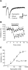
Endogenous κ-receptor agonist dynorphin inhibits glutamate EPSCs. A, EPSCs from a secondary cell in control, in dynorphin (300 nm), and after washout of dynorphin. B, A plot of normalized amplitudes of pooled EPSCs from secondary cells in control (open circles, n = 11) and in nor-BNI (100 nm, filled circles, n = 10). C, A dose-response plot of EPSC inhibition by dynorphin in secondary cells (n = 7-8 cells at each concentration).
Figure 3.
Figure 3.
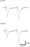
U69593 increases the paired-pulse ratio (PPR) of glutamate EPSCs. Pairs of EPSCs from a primary cell in control and in U69593. Note the increase by U69593 (300 nm) in the PPR (ratio of the second EPSC amplitude over the first).
Figure 4.
Figure 4.
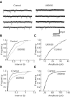
U69593 reduces the frequency of spontaneous glutamate EPSCs. A, Current traces showing spontaneous EPSCs from a primary cell in control and in U69593 (300 nm). B, C, Plots of cumulative distribution of interevent intervals and EPSC amplitudes in control and in U69593 for the cell in A. D, E, Distribution plots for a secondary cell.
Figure 5.
Figure 5.
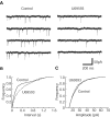
U69593 inhibits the frequency of miniature glutamate EPSCs. Data from a secondary cell. A, Current traces showing miniature EPSCs in control and in U69593 (300 nm). B, C, Plots of cumulative distribution of interevent intervals and miniature EPSC amplitudes in control and in U69593.
Figure 6.
Figure 6.
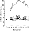
Glutamate receptor antagonists in the NRM block local μ-opioid-induced analgesia. Tail-flick latencies were measured every 2 min before (BL, baseline, average of 6 trials) and after drug microinjections into the NRM and then into the PAG (arrow, time = 0). The cutoff time was 12 sec. Group 1 (open circles): saline in NRM and DAMGO in PAG; group 2 (filled circles): AP-5 + CNQX in NRM and DAMGO in PAG; group 3 (open squares): AP-5 + CNQX in NRM and saline in PAG, n = 5 rats in each group, **p < 0.01 (ANOVA for repeated measures and the Tukey-Kramer test of post hoc analysis).
Figure 7.
Figure 7.
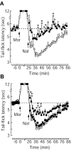
Glutamate receptor antagonists and κ-receptor agonist attenuate opioid abstinence-induced hyperalgesia. After measurements of baseline tail-flick latencies, rats were injected with morphine (2 mg/kg, i.p.) and followed by naloxone (1 mg/kg, i.p.) 26 min later to induce opioid abstinence (withdrawal)-induced hyperalgesia. NRM microinjections were made immediately after the naloxone injection. The dashed line indicates pre-morphine baseline. A, Open circles: saline in NRM (n = 5 rats). Filled circles: AP-5 + CNQX in NRM (n = 6 rats). B, Open circles: same as in A. Filled circles: U69593 in NRM (n = 6 rats). Filled squares: U69593 + nor-BNI in NRM (n = 5 rats). *p < 0.05, **p < 0.01 (ANOVA for repeated measures and the Tukey-Kramer test of post hoc analysis).

References

    1. Ackley MA, Hurley RW, Virnich DE, Hammond DL ( 2001) A cellular mechanism for the antinociceptive effect of a kappa opioid receptor agonist. Pain 91: 377-388. - PubMed
    1. Aimone LD, Gebhart GF ( 1986) Stimulation-produced spinal inhibition from the midbrain in the rat is mediated by an excitatory amino acid neurotransmitter in the medial medulla. J Neurosci 6: 1803-1813. - PMC - PubMed
    1. Cheng HY, Pitcher GM, Laviolette SR, Whishaw IQ, Tong KI, Kockeritz LK, Wada T, Joza NA, Crackower M, Goncalves J, Sarosi I, Woodgett JR, Oliveira-dos-Santos AJ, Ikura M, van der Kooy D, Salter MW, Penninger JM ( 2002) DREAM is a critical transcriptional repressor for pain modulation. Cell 108: 31-43. - PubMed
    1. Coderre TJ, Katz J, Vaccarino AL, Melzack R ( 1993) Contribution of central neuroplasticity to pathological pain: review of clinical and experimental evidence. Pain 52: 259-285. - PubMed
    1. Fields H, Basbaum A ( 1999) Central nervous system mechanisms of pain modulation. In: Textbook of pain, 4E Ed (Wall P, Melzack R, eds). London: Churchill Livingstone.

Publication types

MeSH terms