. 2003 Aug 13;23(19):7269-80.
doi: 10.1523/JNEUROSCI.23-19-07269.2003.

Hypertrophic neuropathies and malignant peripheral nerve sheath tumors in transgenic mice overexpressing glial growth factor beta3 in myelinating Schwann cells

Affiliations

Hypertrophic neuropathies and malignant peripheral nerve sheath tumors in transgenic mice overexpressing glial growth factor beta3 in myelinating Schwann cells

Richard P H Huijbregts et al. J Neurosci. .

Abstract

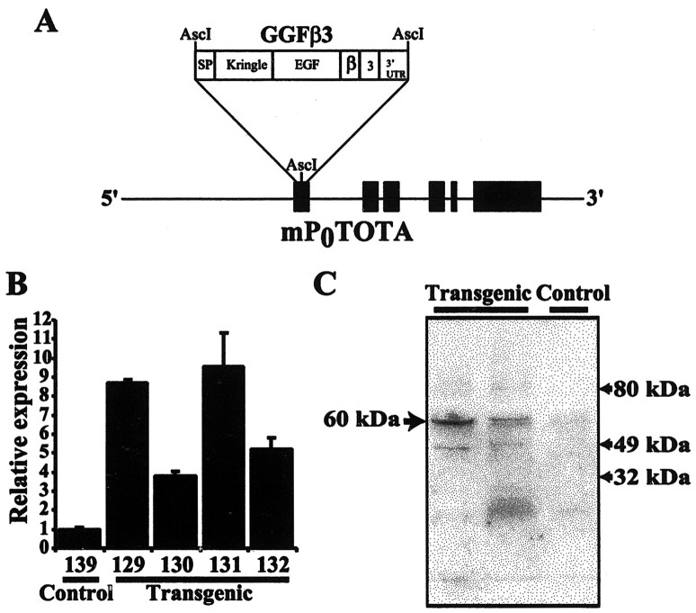
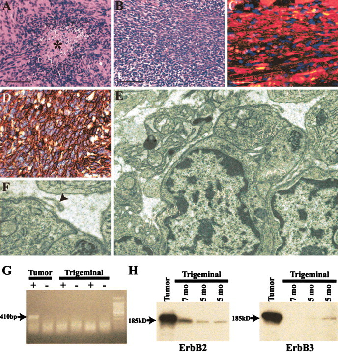
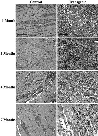
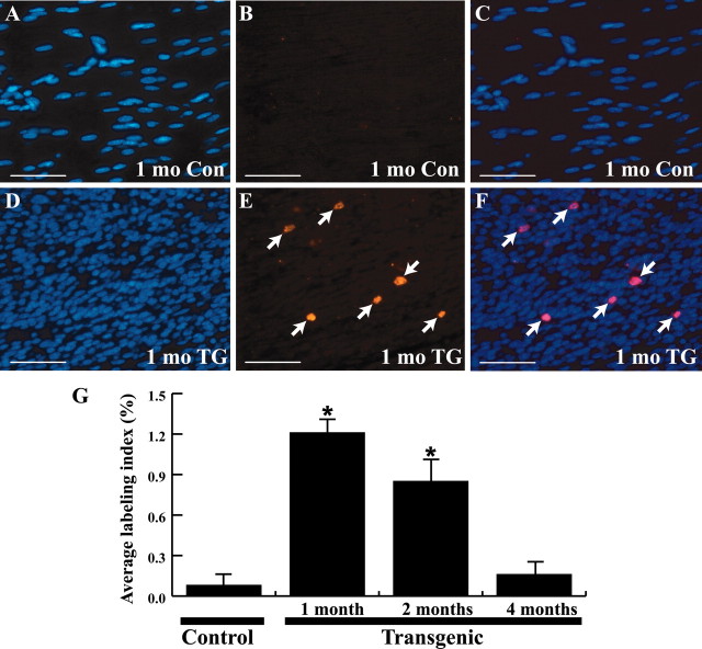
The neuregulin-1 (NRG-1) family of growth and differentiation factors exerts a variety of effects on Schwann cells and their precursors during nervous system development; however, NRG-1 effects on adult Schwann cells are poorly defined. Several lines of evidence suggest that NRG-1 actions on adult Schwann cells are distinct from those observed during development. To test this hypothesis, we generated transgenic mice overexpressing the NRG-1 isoform glial growth factor beta3 (GGFbeta3) in myelinating Schwann cells [protein zero (P0)GGFbeta3 mice]. P0-GGFbeta3 mice develop resting tremors, gait abnormalities, decreased hindlimb strength, and paralysis by approximately 7 months of age. Sciatic nerves from these animals show a hypertrophic neuropathy characterized by demyelination, remyelination, and "onion bulb" formation. Development of this hypertrophic neuropathy is preceded by Schwann cell hyperplasia that is prominent in 1-month-old mice and present but decreased in 2- and 4-month-old animals. P0-GGFbeta3 mice also develop peripheral ganglion-associated malignant peripheral nerve sheath tumors. Motor, sensory, and sympathetic ganglia from 1-, 2-, and 4-month-old P0-GGFbeta3 mice uniformly contain intraganglionic, likely preneoplastic, Schwann cell proliferations. Examination of bromodeoxyuridine incorporation and caspase-3 activation in sciatic nerves and trigeminal ganglia indicates that Schwann cell hyperplasia in P0-GGFbeta3 mice reflects increased proliferation rather than decreased apoptosis. These observations are consistent with the hypothesis that GGFbeta3 induces proliferation of adult Schwann cells and demyelination of peripheral nerve axons. Furthermore, overexpression of this NRG-1 isoform frequently induces neoplastic Schwann cell proliferation within PNS ganglia, suggesting that NRG-1 may contribute to human Schwann cell neoplasia.


Figures

Figure 1.
Figure 1.
Mice carrying the P0-GGFβ3 transgene overexpress GGFβ3 in sciatic nerve. A, Schematic representation of the P0-GGFβ3 transgene in which the expression of a rat GGFβ3 cDNA is directed by 6.0 kb of 5′ flanking sequence and intragenic regulatory elements from the mouse myelin P0 locus contained in mP0TOTA (Feltri et al., 1999). Black boxes indicate exons of the P0 gene, with the structural domains of the GGFβ3 cDNA indicated as white boxes. The GGF domains are as follows: SP, signal peptide; Kringle, kringle domain; EGF, EGF-like domain; β, EGF β-variant domain; 3, 3 variant juxtamembrane domain; and 3′-UTR, the GGFβ3 3′ untranslated region. The P0 start codon in mP0TOTA has been replaced with an AscI site, and the GGFβ3 cDNA has been inserted into this site. B, Semiquantitative RT-PCR analyses of RNA isolated from the sciatic nerves of P0-GGFβ3 mice (line 33) demonstrate that GGFβ3 mRNA is overexpressed in this tissue. Semiquantitative RT-PCR was performed using primers specific for GGFβ3 and the housekeeping gene cyclophilin with RNA isolated from the sciatic nerves of 4-month-old transgenic mice (Transgenic) or age-matched, nontransgenic controls (Control). Signals for GGFβ3 were normalized to cyclophilin levels in the same specimen. GGFβ3/cyclophilin ratios are presented as bars, with SEM indicated for each bar. The GGFβ3/cyclophilin ratio from a nontransgenic mouse (139) is arbitrarily defined as 1, with the level of expression in individual mice normalized to this reference. Numbers under each bar are the identifiers of specific animals. C, Immunoblot analyses of protein from the sciatic nerves of P0-GGFβ3 mice demonstrate that this tissue contains elevated levels of neuregulin-1 protein. Twenty micrograms of sciatic nerve protein from 2-month-old transgenic mice and nontransgenic littermates (line 33) were resolved, blotted, and probed with an antibody specific for the NRG-1 EGF-like common domain [a pan-neuregulin antibody (Carroll et al., 1997)]. The arrow to the left indicates the position of a 60 kDa antigen that is expressed at increased levels in two different transgenic mice (Transgenic) relative to a nontransgenic control (Control). Arrows to the right indicate the positions of size markers.
Figure 2.
Figure 2.
Gait abnormalities and hindlimb paralysis are caused by a hypertrophic neuropathy in P0-GGFβ3 founders 44 and 35. A, Hindlimb paralysis, evident as leg dragging while ambulating, in P0-GGFβ3 founder 44. B-D, Plastic semithin cross sections of sciatic nerves from transgenic founders 35 (B) and 44 (C) compared to an age-matched nontransgenic control (D). These 1 μm sections have been stained with toluidine blue. Numerous onion bulbs are evident in nerves from both founders (e.g., B, arrowhead). Furthermore, numerous large axons, which are thinly myelinated (B, arrows), are present. Occasional actively degenerating axons are also seen (C, arrowheads). Magnification (B-D), 1000×; scale bars (B-D), 25 μm. E, Electron micrograph showing a lipid-laden macrophage in the sciatic nerve from founder 44. Magnification, 6 · 103×. F, Electron micrograph demonstrating a demyelinated large axon in sciatic nerve from founder 44. Magnification, 15 · 103×.
Figure 3.
Figure 3.
A hypertrophic neuropathy whose development is preceded by a period of Schwann cell hyperplasia occurs in mice overexpressing GGFβ3 in myelinating Schwann cells. Light photomicrographs of representative toluidine blue-stained plastic semithin cross sections of sciatic nerves from P0-GGFβ3 mice at 1 (A), 2 (B), and 7 (C) months of age are shown. Arrows in B and C indicate onion bulbs. Arrowheads in C indicate some of the thinly myelinated large axons.
Figure 4.
Figure 4.
A, Representative hematoxylin- and eosin-stained longitudinal sections of sciatic nerve from P0-GGFβ3 transgenic mice (Transgenic) and age-matched nontransgenic controls (Control) at 1, 2, 4, and 7 months of age; ages, indicated to the left of each row, apply both to the images in that row and the adjacent bar graph. Note that sciatic nerves from transgenic animals have higher Schwann cell densities at all ages than seen in age-matched controls. In both transgenic and control mice, however, the number of Schwann cell nuclei per 0.3 mm2 decreases with age. Magnification, 40×; scale bars, 50 μm. B, Bars indicate the average number of Schwann cell nuclei per high-power field at (top to bottom) 1, 2, 4, and 7 months of age in sciatic nerves from P0-GGFβ3 transgenic mice (Transgenic) and age-matched nontransgenic controls (Control). SD is indicated for each bar. Numbers below each bar are animal identifiers. C, Alterations in the average number of Schwann cell nuclei per high-power field (0.3 mm2) with age in P0-GGFβ3 transgenic mice (TG) and nontransgenic littermates (CON). SEM is indicated for each bar. The age of the animals is indicated below each pair of bars.
Figure 5.
Figure 5.
Hyperplastic cells in the sciatic nerves of P0-GGFβ3 mice are associated with Schwann cell markers. A, Immunoreactivity for the Schwann cell marker S100β (red staining) in the sciatic nerve from a 1-month-old P0-GGFβ3 mouse. This preparation has been counterstained with the nuclear marker bisbenzamide to demonstrate the hyperplastic elements. B, C, Immunoreactivity for the basal lamina protein collagen type IV is seen as faint brown staining surrounding Schwann cell units in the sciatic nerve from a 1-month-old nontransgenic mouse (B). In contrast, there is prominent collagen type IV deposition in the sciatic nerve from a 1-month-old P0-GGFβ3 mouse (C), some of which is seen to surround individual hyperplastic cells within this tissue (arrows). In B and C, sections have been counterstained with hematoxylin to highlight nuclei within the sections. Magnification, 63×; scale bars, 50 μm.
Figure 6.
Figure 6.
P0-GGFβ3 transgenic mice develop tumors resembling human malignant peripheral nerve sheath tumors. A, B, Representative sections of trigeminal ganglion-associated tumors from P0-GGFβ3 founders 34 (A) and 33 (B). The asterisk in A indicates a focus of tumor necrosis. Magnification, 40×; scale bar, 50 μm. C, Section of the neoplasm shown in B immunostained for the Schwann cell marker S100β (red staining). This preparation has been counterstained with the nuclear marker bisbenzamide. Magnification, 63×; scalebar 20 μm. D, Section of the neoplasm shown in B immunostained for the basement membrane protein collagen type IV. Collagen type IV immunoreactivity is seen as brown staining that invests individual tumor cells. This preparation has been counterstained with hematoxylin to demonstrate individual tumor cells. Magnification, 63×; scale bar, 20 μm. E, F, Transmission electron micrographs of a neoplasm developing in an offspring of founder 33. Note that individual tumor cells are surrounded by a basal lamina (shown at higher magnification in F), with loops of basal lamina material frequently seen extending away from the tumor cells (F, arrowhead). Magnification: E, 10,000×; F, 40,000×. G, Expression of the P0-GGFβ3 transgene is maintained in the MPNST-like tumors developing in P0-GGFβ3 mice. The transgene mRNA is detected as a 410 bp PCR product in cDNA from the tumor shown in E and F (Tumor) but not in cDNAs from age-matched nontransgenic trigeminal nerves (Trigeminal). The transgene product is not detected in the absence of reverse transcription (- lanes), indicating that the PCR product obtained in these experiments is not derived from contaminating genomic DNA. H, Tumors from P0-GGFβ3 mice express high levels of the NRG-1 receptor subunits erbB2 and erbB3. ErbB2 (left) and erbB3 (right) are detected as 185 kDa immunoreactive species in lysates of the tumor shown in E and F (Tumor) and in trigeminal nerve (Trigeminal) collected from 5- and 7-month-old P0-GGFβ3 mice.
Figure 7.
Figure 7.
The MPNST-like tumors in P0-GGFβ3 transgenic mice likely arise from preneoplastic lesions uniformly present in peripheral ganglia from these mice. Shown are representative hematoxylin- and eosin-stained sections of the trigeminal ganglia from P0-GGFβ3 transgenic mice (Transgenic) and nontransgenic age-matched controls (Control). The ages of these animals are indicated at the left. Magnification, 20×; scale bars, 100 μm.
Figure 8.
Figure 8.
DNA synthesis in sciatic nerves from P0-GGFβ3 transgenic and wild-type mice. A-C, Representative section from a 1-month-old wild-type control (1 mo Con) mouse. D-F, Representative section from a 1-month-old P0-GGFβ3 transgenic (1 mo TG) mouse. Left column, Staining for the nuclear dye bisbenzamide. Middle column, Immunostaining for incorporated BrdU. Right column, Merging of images in the left and middle columns. Arrows in E and F indicate BrdU-positive Schwann cell nuclei. Scale bars, 50 μm. G, BrdU labeling indices determined in the sciatic nerves of P0-GGFβ3 transgenic mice and age-matched wild-type controls. The percentage of Schwann cell nuclei labeled with BrdU is indicated on the left, with bars indicating the average percentage labeled and the SEM. The age of the animals is indicated below the bars. Sections of jejunum from each animal were also immunostained (data not shown); BrdU incorporation in the proliferative zones of the crypts of Lieberkuhn served as a positive control for BrdU delivery. BrdU labeling indices for sciatic nerves from P0-GGFβ3 transgenic mice and age-matched wild-type controls were compared using one-way ANOVA, followed by Tukey'sposthoctest, withp < 0.05 considered statistically significant. *p << 0.001 relative to nontransgenic controls.

Similar articles

Cited by

References

    1. Adlkofer K, Lai C ( 2000) Role of neuregulins in glial cell development. Glia 29: 104-111. - PubMed
    1. Anderson LM, Hagiwara A, Kovatch RM, Rehm S, Rice JM ( 1989) Transplacental initiation of liver, lung, neurogenic and connective tissue tumors by N-nitroso compounds in mice. Fund Appl Toxicol 12: 604-620. - PubMed
    1. Arpornchayanon O, Hirota T, Itabashi M, Nakajima T, Fukuma H, Beppu Y, Nishikawa K ( 1984) Malignant peripheral nerve tumors: a clinicopathological and electron microscopic study. Jpn J Clin Oncol 14: 57-74. - PubMed
    1. Asbury AK ( 1967) Schwann cell proliferation in developing mouse sciatic nerve: a radioautographic study. J Cell Biol 34: 735-743. - PMC - PubMed
    1. Baron-Van Evercooren A, Gansmuller A, Gumpel M, Baumann N, Kleinman HK ( 1986) Schwann cell differentiation in vitro: extracellular matrix deposition and interaction. Dev Neurosci 8: 182-196. - PubMed

Publication types

MeSH terms