. 2003 Aug 13;23(19):7288-97.
doi: 10.1523/JNEUROSCI.23-19-07288.2003.

Presynaptic and postsynaptic mechanisms of a novel form of homosynaptic potentiation at aplysia sensory-motor neuron synapses

Affiliations

Presynaptic and postsynaptic mechanisms of a novel form of homosynaptic potentiation at aplysia sensory-motor neuron synapses

Iksung Jin et al. J Neurosci. .

Abstract

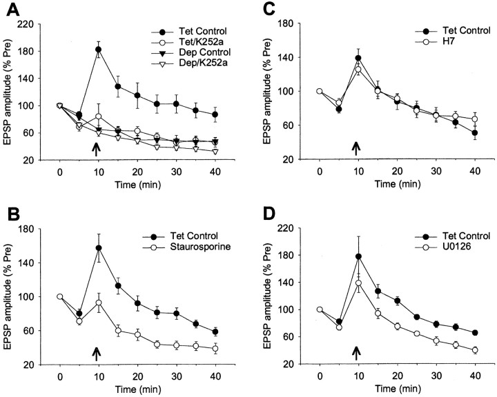
Previous studies have shown that homosynaptic potentiation produced by rather mild tetanic stimulation (20 Hz, 2 sec) at Aplysia sensory-motor neuron synapses in isolated cell culture involves both presynaptic and postsynaptic Ca2+ (Bao et al., 1997). We have now investigated the sources of Ca2+ and some of its downstream targets. Although the potentiation lasts >30 min, it does not require Ca2+ influx through either NMDA receptor channels or L-type Ca2+ channels. Rather, the potentiation involves metabotropic receptors and intracellular Ca2+ release from both postsynaptic IP3-sensitive and presynaptic ryanodine-sensitive stores. In addition, it involves protein kinases, including both presynaptic and postsynaptic CamKII and probably MAP kinase. Finally, it does not require transsynaptic signaling by nitric oxide but it may involve AMPA receptor insertion. The potentiation, thus, shares components of the mechanisms of post-tetanic potentiation, NMDA- and mGluR-dependent long-term potentiation, and even long-term depression, but is not identical to any of them. These results are consistent with the more general idea that there is a molecular alphabet of basic components that can be combined in various ways to create novel as well as known types of plasticity.


Figures

Figure 1.
Figure 1.
The experimental preparation and protocol. A, Micrograph of an Aplysia gill motor neuron (L7) isolated from the abdominal ganglion of a juvenile animal and cocultured with two sensory neurons (SN) from the pleural ganglion of an adult animal. One of the sensory neurons was stimulated with an extracellular electrode pressed against the cell body, and the evoked EPSP was recorded with a sharp intracellular electrode in the motor neuron. B, General protocol. After 30 min of rest or drug incubation, the EPSP was tested once every 5 min for 40 min. Tetanic stimulation (20 Hz, 2 sec) was delivered to the sensory neuron 1 min before the third test. C, Examples of the PSPs in a representative potentiation experiment and in a control experiment without tetanic stimulation. D, The average change in the PSP in all of the no-drug potentiation experiments (n = 119) and test-alone (homosynaptic depression) control experiments (n = 37) in this study. The data have been normalized to the value on the pretest in each experiment. The arrow indicates the time of tetanic stimulation, and the error bars indicate the SEM. There was no difference between experiments with and without DMSO, which have been pooled.
Figure 2.
Figure 2.
Homosynaptic potentiation does not require Ca2+ influx through NMDA receptor channels or voltage-dependent Ca2+ channels. A, Homosynaptic potentiation is not affected by an antagonist of NMDA-type glutamate receptors, APV (50 μm). B, Homosynaptic potentiation is not affected by an inhibitor of L-type Ca2+ channels, nitrendipine (10 μm). C, Homosynaptic potentiation is also not affected by reducing extracellular Ca2+ from 10 to 2 mm. D, Homosynaptic potentiation is reduced by an antagonist of type I metabotropic glutamate receptors, LY367385 (300 μm).
Figure 3.
Figure 3.
Homosynaptic potentiation involves Ca 2+ release from both IP3- and ryanodine-sensitive intracellular stores. A, Examples of the PSPs in a representative potentiation experiment and a depression control experiment during perfusion with thapsigargin (10 μm), which depletes intracellular Ca2+ stores. B, Average results from experiments like these shown in A. Homosynaptic potentiation is greatly reduced by thapsigargin, compared with the no drug control experiments. Thapsigargin also tended to increase homosynaptic depression on the second test, 5 min after the first PSP, but had no significant effect on the depression from 10 to 40 min. There was a significant drug × tetanus interaction (F(1, 32) = 13.83; p < 0.001) in a three-way ANOVA with one repeated measure (time) for the data from 10 to 40 min. C, Homosynaptic potentiation is greatly reduced by a cell permeable inhibitor of IP3 receptors, 2APB (10 μm). D, Homosynaptic potentiation is reduced by prolonged application of a relatively high concentration (100 μm) of ryanodine, which blocks ryanodine receptors. Inset, Brief application of a relatively low concentration (1 μm) of ryanodine (which activates ryanodine receptors) after the first PSP produced potentiation of the second PSP. The average amplitudes of the PSPs on the first test were 20.4 mV (control, n = 9) and 6.8 mV (1 μm ryanodine, n = 6); not significantly different.
Figure 4.
Figure 4.
Homosynaptic potentiation involves both presynaptic and postsynaptic Ca2+ release, and short-term plasticity also involves presynaptic Ca2+ release. A, Homosynaptic potentiation is significantly reduced by injection of a cell impermeable antagonist of IP3 receptors (heparin, 2 mg/ml in the electrode) but not a cell impermeable antagonist of ryanodine receptors (8NcADPR, 20 μm in the electrode) into the motor neuron. B, Homosynaptic potentiation is significantly reduced by injection of 8NcADPR, but not heparin, into the sensory neuron. C, Short-term depression during the tetanus is increased by thapsigargin or a high concentration of ryanodine, but not by 2APB. The data are from the same experiments as Figure 3. To get an overall index of plasticity during the 2 sec tetanus, we measured the total area under the tetanus (in millivolts × milliseconds) normalized to the amplitude of the first PSP in the tetanus (in millivolts) in each experiment. D, Short-term depression during the tetanus is increased by presynaptic 8NcADPR but not by postsynaptic 8NcADPR or either presynaptic or postsynaptic heparin. The data are from the same experiments as A and B.
Figure 5.
Figure 5.
Homosynaptic potentiation involves protein kinases, probably including MAP kinase. A, Homosynaptic potentiation is greatly reduced by a general inhibitor of protein kinases, K252a (200 nm). K252a also increased homosynaptic depression on the second test, 5 min after the first PSP, but had no significant effect on the depression from 10 to 40 min. There was a significant drug × tetanus interaction (F(1,21) = 16.68; p < 0.001) in a three-way ANOVA with one repeated measure (time) for the data from 10 to 40 min. B, Homosynaptic potentiation is also reduced by another general inhibitor of protein kinases, staurosporine (50 nm). C, Homosynaptic potentiation is not reduced by a third general inhibitor of protein kinases, H7 (100 μm). D, Homosynaptic potentiation is reduced by an inhibitor of MAP kinase, U0126 (10 μm).
Figure 6.
Figure 6.
Homosynaptic potentiation involves both presynaptic and postsynaptic CamKII. A, Homosynaptic potentiation is greatly reduced by a more specific inhibitor of CamKII, KN93 (5 μm). KN93 also tended to increase homosynaptic depression on the second test, 5 min after the first PSP, but had no significant effect on the depression from 10 to 40 min. There was a significant drug × tetanus interaction (F(1,23) = 10.67; p < 0.01) in a three-way ANOVA with one repeated measure (time) for the data from 10 to 40 min. B, Homosynaptic potentiation is also reduced by an inhibitor of calmodulin, calmidazolium (10 μm). C, Homosynaptic potentiation is reduced by injection of a peptide inhibitor of CamKII, CamKII 281-309 (300 μm in the electrode) into the motor neuron. D, Homosynaptic potentiation is also reduced by injection of CamKII 281-309 into the sensory neuron.
Figure 7.
Figure 7.
Homosynaptic potentiation does not require NO but may involve AMPA receptor insertion. A, Homosynaptic potentiation is not reduced by an inhibitor of NOS, Nω-nitro-l-arginine (500 μm). B, Homosynaptic potentiation is also not reduced by a scavenger of NO, oxymyoglobin (10 μm), compared with the inactive form, metmyoglobin. C, Homosynaptic potentiation is greatly reduced by an antagonist of AMPA-type glutamate receptors, DNQX (5 μm). DNQX also increased homosynaptic depression on the second test, 5 min after the first PSP, but had no significant effect on the depression from 10 to 40 min. There was a significant drug × tetanus interaction (F(1,22) = 10.68; p < 0.01) in a three-way ANOVA with one repeated measure (time) for the data from 10 to 40 min. D, Homosynaptic potentiation is also reduced by injection of the light chain of botulinum toxin type B (0.5 μm in the electrode), which blocks exocytocis, into the motor neuron.

References

    1. Angers A, Fioravante D, Chin J, Cleary LJ, Bean AJ, Byrne JH ( 2002) Serotonin stimulates phosphorylation of Aplysia synapsin and alters its sub-cellular distribution in sensory neurons. J Neurosci 22: 5412-5422. - PMC - PubMed
    1. Antonov I, Antonova I, Minnal A, Hawkins RD ( 2001) Possible interaction of pre- and postsynaptic mechanisms during classical conditioning in Aplysia Soc Neurosci Abstr 27: 954.15.
    1. Antonov I, Antonova I, Kandel ER, Hawkins RD ( 2003) Activity-dependent presynaptic facilitation and Hebbian LTP are both required and interact during classical conditioning in Aplysia Neuron 37: 135-147. - PubMed
    1. Anwyl R, Lee WL, Rowan M ( 1988) The role of calcium in short-term potentiation in the rat hippocampal slice. Brain Res 459: 192-195. - PubMed
    1. Bao JX, Kandel ER, Hawkins RD ( 1997) Involvement of pre- and postsynaptic mechanisms in posttetanic potentiation at Aplysia synapses. Science 275: 969-973. - PubMed

Publication types

MeSH terms

Substances

LinkOut - more resources