. 2003 Aug 13;23(19):7415-25.
doi: 10.1523/JNEUROSCI.23-19-07415.2003.

Freud-1: A neuronal calcium-regulated repressor of the 5-HT1A receptor gene

Affiliations

Freud-1: A neuronal calcium-regulated repressor of the 5-HT1A receptor gene

Xiao-Ming Ou et al. J Neurosci. .

Abstract

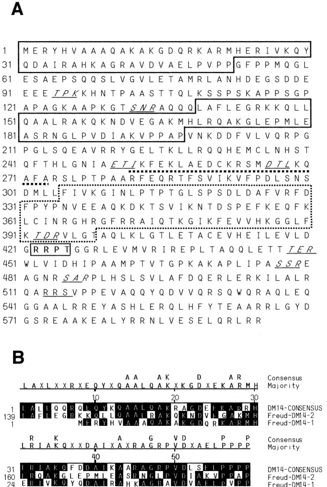
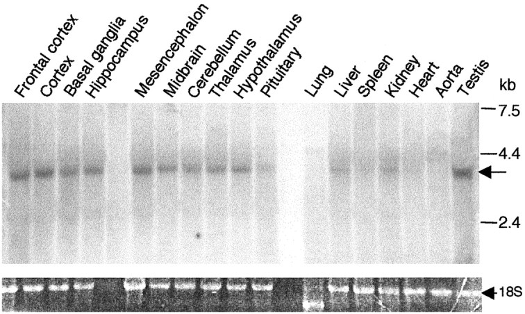
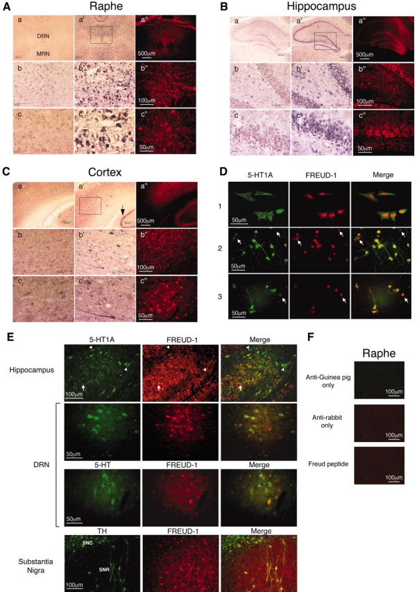
Altered regulation of 5-HT1A receptors is implicated in mood disorders such as anxiety and major depression. To provide insight into its transcriptional regulation, we previously identified a novel DNA element [14 bp 5'-repressor element (FRE)] of the 5-HT1A receptor gene that mediates repression in neuronal and non-neuronal cells (Ou et al., 2000). We have now cloned a novel DNA binding protein [five' repressor element under dual repression binding protein-1 (Freud-1)] that binds to FRE to mediate repression of the 5-HT1A receptor or heterologous promoters. Freud-1 is evolutionarily conserved and contains two DM-14 basic repeats, a predicted helix-loop-helix DNA binding domain, and a protein kinase C conserved region 2 (C2)/calcium-dependent lipid binding (CalB) calcium/phospholipid binding domain. An intact CalB domain was required for Freud-1-mediated repression. In serotonergic raphe cells, overexpression of Freud-1 repressed the 5-HT1A promoter and decreased 5-HT1A receptor protein levels, whereas transfection of antisense to Freud-1 derepressed the 5-HT1A gene and increased 5-HT1A receptor protein expression. Calcium-dependent signaling blocked Freud-1-FRE binding and derepressed the 5-HT1A promoter. Treatment with inhibitors of calmodulin or CAM-dependent protein kinase reversed calcium-mediated inhibition of Freud-1. Freud-1 RNA and protein were present in raphe nuclei, hippocampus, cortex, and hypothalamus, and Freud-1 protein was colocalized with 5-HT1A receptors, suggesting its importance in regulating 5-HT1A receptors in vivo. Thus, Freud-1 represents a novel calcium-regulated repressor that negatively regulates basal 5-HT1A receptor expression in neurons and may play a role in the altered regulation of 5-HT1A receptors associated with anxiety or major depression.


Figures

Figure 1.
Figure 1.
Protein structure of Freud-1. A, Predicted structural motifs in Freud-1 are shown. Indicated are consensus sequences for DM-14 (boxed) and C2 domains (dashed box), helix-loop-helix DNA binding domain (underlined), cAMP phosphorylation site (bold and outlined), and PKC phosphorylation sites (italic and underlined). B, Alignment of Freud-1 DM-14 sequence with consensus sequence.
Figure 2.
Figure 2.
Specific binding of Freud-1 to 5-HT1A-FRE requires an intact CalB motif. The sequences of oligonucleotides for DRE (31 bp), FRE (14 bp), and TRE (12 bp) are indicated. A, Recombinant Freud-1-DRE binding was assessed by EMSA using 5-HT1A-DRE (31 bp) as a probe incubated with 4 μg of purified Freud-1 (lanes 1-6) or without (lane 7) as indicated. A single specific species (band 1) was retarded in the presence of 10 μg of anti-Freud-1 antibody (band 2, lane 4). The retarded complex was blocked by coincubation with 4 μg of peptide antigen (lane 6). Double-stranded unlabeled competitor oligonucleotides (at 100-fold molar excess) were included as indicated. FRE was sufficient to compete for Freud-1-DRE complex. B, Recombinant Freud-1-FRE binding: 4 μg of purified Freud-1 interacted specifically with FRE (band 1, lane 1). Freud-1-FRE interaction was abolished by preincubation with 100-fold unlabeled FRE (lane 2) but not by TRE (lane 3). C, Recombinant mutated Freud-1-DRE binding: EMSA using DRE as a probe incubated with 8 μg of purified Freud-1 with a disrupted CalB domain (Freud-1 del) alone (lane 1) or with unlabeled 14 bp FRE (lane 2) as indicated. A disrupted CalB motif reduced affinity of Freud-1 for the DRE. D, Presence of Freud-1 in nuclear extracts of L6 and RN46A cells. EMSA using the 31 bp DRE as a probe incubated without (lane 1) or with nuclear extracts from L6 (lanes 2, 3) or RN46A cells (lanes 4, 5) as indicated. The higher mobility complex (band 1) was displaced rather than retarded in the presence of anti-Freud-1 antibody in extracts from both cell lines (lanes 3 and 5, respectively). The second protein/DRE complex present in L6 cells (band 2) was not affected by Freud-1 antibody (lane 3) and represents an additional unknown repressor.
Figure 3.
Figure 3.
Freud-1 represses basal 5-HT1A receptor expression in raphe RN46A cells via FRE. Luciferase activity was normalized to that of β-galactosidase and is expressed as relative light units and normalized to control. *p < 0.05 compared with control by t test. A, CalB-dependent Freud-1 repression. The FRE-containing -2300 5-HT1A-luciferase was transiently cotransfected in L6 myoblast or raphe RN46A cells with vector (pcDNA3, Control), Freud-1, or Freud-1 del mutant of the C2 (CalB) domain. Above, A Western blot of 30 μg of nuclear extracts was probed with anti-Flag to show equal expression of Flag-Freud-1 or Flag-Freud-1 del in these experiments. B, FRE dependence of Freud-1 repression. Transfections as above were done with FRE-inactivated mutant 2300m1, which was insensitive to Freud-1. C, FRE-dependent repression of SV40 promoter by Freud-1. The 31 bp DRE (DRE/SV40) or the FRE-mutant DRE (DREmut/SV40) was placed upstream of the SV40 promoter and cotransfected with vector (pcDNA3) or Freud-1 expression construct. D, E, Freud-1 inhibits transcriptional activity of the 5-HT1A receptor gene. L6 myoblast or raphe RN46A cells were transiently cotransfected with the DRE-containing (-1590luc) 5-HT1A-luciferase reporter or vector (pcDNA3, Control), and sense (D) or antisense (E) Freud-1 expression vectors. Freud-1 protein expression in each cell line after transfection was detected by Western blot using specific anti-Freud antibody (shown above). β-Actin immunoreactivity was tested to confirm equal loading. F, G, Freud-1 protein inhibits 5-HT1A receptor expression in raphe RN46A cells. F, Proteins prepared from RN46A cells transfected with Freud-1 sense or antisense expression vectors and differentiated for 72 hr were subjected to anti-5-HT1A receptor immunoblotting. β-Actin immunoreactivity was tested to confirm equal loading. Shown is a representative blot of three independent experiments. The relative intensity was quantified by an Automated Digitizing System (UN-SCAN-IT, Silk Scientific Inc.). Data represent the mean ± SE of three independent experiments. G, RN46A cells cotransfected with 1 μg of Red Fluorescent Protein (DsRed) and 5 μg of Freud-1 sense or antisense expression plasmids as indicated were stained for 5-HT1A immunoreactivity (see Materials and Methods). Arrows indicate representative DsRed-positive cells that express either high or low 5-HT1A immunoreactivity (for antisense or sense Freud-1, respectively) compared with cells not transfected with DsRed.
Figure 4.
Figure 4.
CAM kinase attenuates Freud-1-mediated repression. A, Ca2+ and ATP in combination interfere with Freud-1/DRE interaction. EMSA using the 31 bp DRE as a probe incubated with nuclear extracts from RN46A (lanes 1-4) or L6 cells (lanes 5-8) as indicated. CaCl2 (lanes 2, 6), ATP (lanes 3, 7), or both (lanes 4, 8) were added into incubation buffer for 20 min before incubation with DRE probe. B, C, CAM kinase activation in nuclear extracts interferes with Freud-1-DRE binding. EMSA using DRE as a probe and nuclear extracts from RN46A cells (B) and L6 cells (C) are shown. Both CaCl2 and ATP were added into the incubation before adding probe (lanes 2-7). Treatment was with 10 μm (except 50 nm CalC) KN92 (negative control for KN93; lane 3), KN93 (CAM kinase inhibitor; lane 4), CalC (PKC inhibitor; lane 5), H-8 (PKA inhibitor, lane 6), and CMZ (CAM inhibitor; lane 7), as indicated. In L6 cells (C), CAM or CAMK inhibitors rescued only the Freud-1-containing species (band 1). D, Calcium signaling enhances 5-HT1A promoter activity in a Freud-1-FRE-dependent manner. 5-HT1A promoter-luciferase reporter constructs containing FRE (-1590 or -2300) or lacking FRE (-1519, -2300m1) were transfected into RN46A cells. Twenty-four hours after transfection, the cells were treated with 40 mm KCl or with 40 mm KCl and 1 μm ionomycin without or with 10 μm KN92, KN93, or CMZ in the medium for 16 hr. Luciferase activity is expressed as relative light units normalized to control (untreated) samples. *p < 0.05 in comparison with control. Note that calcium mobilizing agents had no effect in FRE-lacking reporter constructs (-1519 or -2300m1) and were blocked by CAM or CAMK inhibitors.
Figure 5.
Figure 5.
Tissue distribution of Freud-1 RNA expression. Freud-1 mRNA expression in rat tissues. RNA prepared from 17 different rat tissues was used for Northern blot analysis. The position of molecular mass markers is shown on the right. An arrow indicates hybridization to Freud-1 probe. Ethidium bromide-stained 18S RNA was used as a loading control.
Figure 6.
Figure 6.
Colocalization of Freud-1 with 5-HT1A receptor. A-C, Coronal brain sections were hybridized with sense (a-c) or antisense (a-c′) digoxigenin-labeled Freud-1 oligonucleotides and stained as described in Materials and Methods or were assayed for Freud-1 immunoreactivity (a-c″). Boxed regions are displayed at successively higher magnification in a-c, a-c′, and a-c″. Scale bars: a, a, a″, 500 μm; b, b, b″, 100 μm; c, c, c″, 50 μm. Freud-1 staining was observed in the following regions. A, Raphe nuclei. Staining was most intense in cells of the dorsal raphe nucleus (DRN) but also present in the medial raphe nucleus (MRN) and raphe magnus (data not shown). The dorsal raphe nucleus is magnified showing cytoplasmic Freud-1 RNA and nuclear Freud-1 protein. B, Hippocampus. Staining is prominent in pyramidal cells of CA1, CA3, and the dentate gyrus. Dentate gyrus cells are magnified to show pyramidal cell morphology. C, Primary sensory cortex. The arrow indicates hippocampus for comparison. D, Nuclear localization of Freud-1. Colocalization of Freud-1 (red) and 5-HT1A receptors (green) in raphe RN46A cells (1), and primary cultures of embryonic cortical (2) and hippocampal (3) cells. Arrows indicate cells expressing high levels of Freud-1 and low levels of 5-HT1A receptor. E, Colocalization studies. Dual immunofluorescence was used to detect colocalization of Freud-1 with various markers. Freud-1 was colocalized with 5-HT1A receptor staining in sections from hippocampus and dorsal raphe nucleus (DRN), as indicated. In DRN, Freud-1 was also colocalized with staining for 5-HT, a marker of serotonergic neurons. In the substantia nigra pars compacta (SNC) or especially pars reticulata (SNR), Freud-1 was colocalized with TH, a marker for dopaminergic neurons. Arrowheads indicate cells expressing low Freud-1 and high 5-HT1A receptor levels. F, Specificity of 5-HT1A and Freud-1 staining. Raphe tissue sections that were incubated with anti-guinea pig or anti-rabbit secondary antibodies alone or in the presence of 100 μg of Freud-1 peptide antigen displayed only background immunoreactivity.

References

    1. Abdouh M, Storring JM, Riad M, Paquette Y, Albert PR, Drobetsky E, Kouassi E ( 2001) Transcriptional mechanisms for induction of 5-HT1A receptor mRNA and protein in activated B and T lymphocytes. J Biol Chem 276: 4382-4388. - PubMed
    1. Albert PR, Tashjian Jr AH ( 1986) Ionomycin acts as an ionophore to release TRH-regulated Ca 2+ stores from GH4C1 cells. Am J Physiol 251: C887-891. - PubMed
    1. Albert PR, Zhou QY, Van Tol HH, Bunzow JR, Civelli O ( 1990) Cloning, functional expression, and mRNA tissue distribution of the rat 5-hydroxy-tryptamine1A receptor gene. J Biol Chem 265: 5825-5832. - PubMed
    1. Albert PR, Lembo P, Storring JM, Charest A, Saucier C ( 1996) The 5-HT1A receptor: signaling, desensitization, and gene transcription. Neuropsychopharmacology 14: 19-25. - PubMed
    1. Albert PR, Sajedi N, Lemonde S, Ghahremani MH ( 1999) Constitutive G(i2)-dependent activation of adenylyl cyclase type II by the 5-HT1A receptor. Inhibition by anxiolytic partial agonists. J Biol Chem 274: 35469-35474. - PubMed

Publication types

MeSH terms