. 2003 Sep 30;100 Suppl 1(Suppl 1):11861-5.
doi: 10.1073/pnas.1834205100. Epub 2003 Aug 13.

Ingestion of bacterially expressed double-stranded RNA inhibits gene expression in planarians

Affiliations

Ingestion of bacterially expressed double-stranded RNA inhibits gene expression in planarians

Phillip A Newmark et al. Proc Natl Acad Sci U S A. .

Abstract

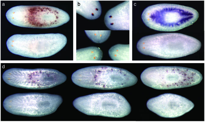
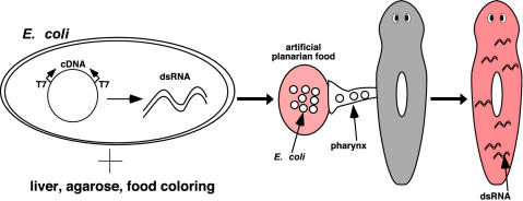
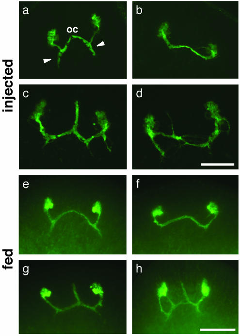
Freshwater planarian flatworms are capable of regenerating complete organisms from tiny fragments of their bodies; the basis for this regenerative prowess is an experimentally accessible stem cell population that is present in the adult planarian. The study of these organisms, classic experimental models for investigating metazoan regeneration, has been revitalized by the application of modern molecular biological approaches. The identification of thousands of unique planarian ESTs, coupled with large-scale whole-mount in situ hybridization screens, and the ability to inhibit planarian gene expression through double-stranded RNA-mediated genetic interference, provide a wealth of tools for studying the molecular mechanisms that regulate tissue regeneration and stem cell biology in these organisms. Here we show that, as in Caenorhabditis elegans, ingestion of bacterially expressed double-stranded RNA can inhibit gene expression in planarians. This inhibition persists throughout the process of regeneration, allowing phenotypes with disrupted regenerative patterning to be identified. These results pave the way for large-scale screens for genes involved in regenerative processes.


Figures

Fig. 2.
Fig. 2.
Inhibition of gene expression in planarians by ingestion of bacterially expressed dsRNA. (a) Specific inhibition of a centrally expressed MP. Both animals were fed bacteria expressing a construct targeting an astacin-like MP (B10). One animal (Upper) was hybridized to detect the transcript encoded by another centrally expressed MP (collagenase-like B2; positive signal in 10/10), whereas another animal (Lower) was hybridized to detect B10 (no detectable expression in 32/36; significantly reduced expression in 3/36). When MP B2 was targeted, and animals were fixed and hybridized to detect B2 transcripts, no signal was detected in 39/47 samples; 8/47 showed a strong reduction of transcript (data not shown). (b) Inhibition of photoreceptor expression of arrestin E30. (Upper) The animals were fed bacteria targeting a gut-specific gene; photoreceptor expression of E30 is normal (7/7). (Lower) The animals were fed a construct targeting photoreceptor-specific arrestin (reduction observed in 17/17). (c) Inhibition of expression in the gastrovascular system. Hybridization with gastrovascular-specific probe D14.38. (Upper) The animal was fed a construct targeting a photo-receptor-specific gene (4/4 positive). (Lower) The animal was fed a construct targeting D14.38 (no signal in 44/44 worms). (d) Inhibition persists throughout the regenerative process. (Upper) The animals were fed a control bacterial strain, containing the vector alone. (Lower) The animals were fed bacteria targeting astacin-like MP B10. Three days after feeding, planarians were cut into three parts and allowed to regenerate for 9 days [12 days after the last feeding; animals are regenerated from head fragments (Left), trunk fragments (Center), and tail fragments (Right)]. The control worms reestablished expression of B10 (positive signal in 6/6 treated worms), whereas in the experimental worms B10 expression was inhibited below detectable levels (no detectable expression in 14/14 worms).
Fig. 1.
Fig. 1.
Method for feeding bacteria expressing dsRNA to planarians. dsRNA synthesis is induced in E. coli by using a vector developed for use in C. elegans (15) that contains two T7 RNA polymerase promoters flanking the cDNA sequence of interest. After induction of dsRNA synthesis, bacteria are mixed with homogenized liver (normal planarian food), ultra-low gelling temperature agarose, and red food coloring (for visualization of eating). This mixture solidifies, and animals that ingest this artificial food become red and are exposed to the dsRNA.
Fig. 3.
Fig. 3.
Introduction of H.108.3a dsRNA by injection (a-d) or feeding (e-h) results in similar defects in axonal guidance after regeneration. (a) Control injections; 16/18 regenerated animals display normal axonal trajectories; 2/18 show minor defects distinct from those shown below. (b-d) Phenotypes observed after injection of H.108.3a dsRNA. (b) Regenerated photoreceptor neurons failed to extend axons posteriorly from the optic chiasmata toward the visual center of the brain (8/28 animals). (c) Regenerated photoreceptor neurons had abnormal anterior projections from the optic chiasmata (11/28 animals). (d) Regenerated photoreceptors had ectopic projections or patterning defects (9/28 animals). (e) Control RNAi by feeding; animals were fed bacteria expressing dsRNA from the C. elegans unc-22 gene. After amputation and regeneration, 0/40 animals displayed any abnormalities in axon path-finding. (f-h) Phenotypes observed after feeding of bacterially expressed H.108.3a dsRNA. (f) Regenerated photoreceptor neurons failed to extend axons posteriorly from the optic chiasmata (18/48 animals). (g) Regenerated photoreceptors had abnormal anterior projections from the optic chiasmata (10/48 animals). (h) Regenerated photoreceptors had ectopic projections or patterning defects (4/48). The remainder (16/48) had no observable defects in the pattern of axonal projections (data not shown). oc, optic chiasmata. Arrowheads, posterior axon projections from optic chiasmata to brain. The view is anterior to the top in all frames. (Scale bars: a-d, 100 μm; e-h, 125 μm.)

References

    1. Sánchez Alvarado, A. (2000) BioEssays 22, 578-590. - PubMed
    1. Brøndsted, H. V. (1969) Planarian Regeneration (Pergamon, London).
    1. Agata, K. & Watanabe, K. (1999) Semin. Cell Dev. Biol. 10, 377-383. - PubMed
    1. Newmark, P. A. & Sánchez Alvarado, A. (2002) Nat. Rev. Genet. 3, 210-219. - PubMed
    1. Baguñà, J., Saló, E. & Auladell, C. (1989) Development (Cambridge, U.K.) 107, 77-86.

Publication types

LinkOut - more resources