

Evaluation of aid to diagnosis of pigmented skin lesions in general practice: controlled trial randomised by practice
- PMID: 12919990
- PMCID: PMC175808
- DOI: 10.1136/bmj.327.7411.375
Evaluation of aid to diagnosis of pigmented skin lesions in general practice: controlled trial randomised by practice
Abstract
Objectives: To determine whether an aid to the diagnosis of pigmented skin lesions reduces the ratio of benign lesions to melanomas excised in general practice.
Design: Controlled trial randomised by practice.
Setting: General practices in Perth, Western Australia.
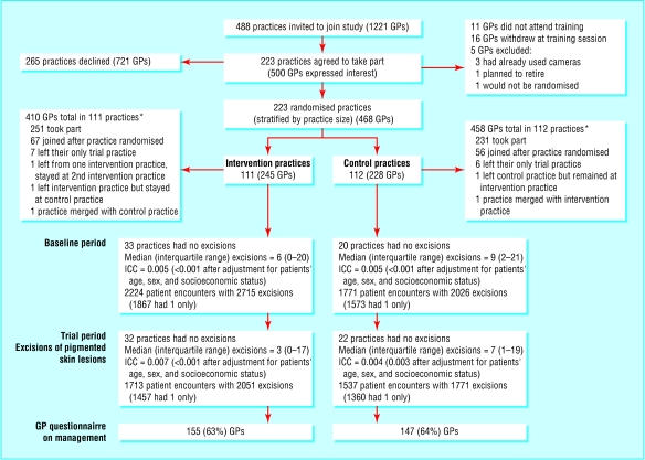
Participants: 468 general practitioners in 223 practices.
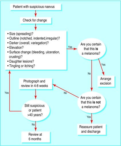
Interventions: Intervention practices were given an algorithm and instant camera to assist with the diagnosis of pigmented skin lesions. All practices were given national guidelines on managing melanoma.
Main outcome measures: Ratio of benign pigmented lesions to melanomas excised. Analyses conducted with and without inclusion of seborrhoeic keratoses.
Results: At baseline the ratios of benign to malignant lesions were lower in the intervention group than in the control group. During the trial period the ratios were higher in the intervention group (19:1 v 17:1 without seborrhoeic keratoses and 29:1 v 26:1 with seborrhoeic keratoses). After adjustment for patients' age, sex, and socioeconomic status, the ratio was 1.02 times higher (95% confidence interval 0.68 to 1.51, P = 0.94) in the intervention group when seborrhoeic keratoses were not included and 1.03 times higher (0.71 to 1.50, P = 0.88) when seborrhoeic keratoses were included. General practitioners in the intervention group were less likely than those in the control group to excise the most recent pigmented skin lesion they managed (22% v 48%, P < 0.001) and to refer the patient to a specialist (16% v 27%, P = 0.06).
Conclusions: Provision of the algorithm and camera did not decrease the ratio of benign pigmented skin lesions to melanomas excised by general practitioners.
Figures
Comment in
-
Diagnosing pigmented skin lesions in general practice: objective assessment of skin lesions is possible.BMJ. 2003 Nov 15;327(7424):1167; author reply 1167. doi: 10.1136/bmj.327.7424.1167-b. BMJ. 2003. PMID: 14615353 Free PMC article. No abstract available.
References
-
- Shaw HM, Balch CM, Soong SJ, Milton GW, McCarthy WH. Prognostic histopathological factors in malignant melanoma. Pathology 1985;17: 271-4. - PubMed
-
- Burton RC, Howe C, Adamson L, Reid AL, Hersey P, Watson A, et al. General practitioner screening for melanoma: sensitivity, specificity, and effect of training. J Med Screen 1998;5: 156-61. - PubMed
-
- Burton RC, Coates MS, Hersey P, Roberts G, Chetty MP, Chen S, et al. An analysis of a melanoma epidemic. Int J Cancer 1993;55: 765-70. - PubMed
-
- Del Mar C, Green A, Cooney T, Cutbush K, Lawrie S, Adkins G. Melanocytic lesions excised from the skin: what percentage are malignant? Aust J Public Health 1994;18: 221-3. - PubMed
Publication types
MeSH terms
LinkOut - more resources
Full Text Sources
Medical