. 2003 Sep 2;100(18):10388-92.
doi: 10.1073/pnas.1434299100. Epub 2003 Aug 14.

B7x: a widely expressed B7 family member that inhibits T cell activation

Affiliations

B7x: a widely expressed B7 family member that inhibits T cell activation

Xingxing Zang et al. Proc Natl Acad Sci U S A. .

Abstract

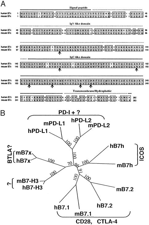
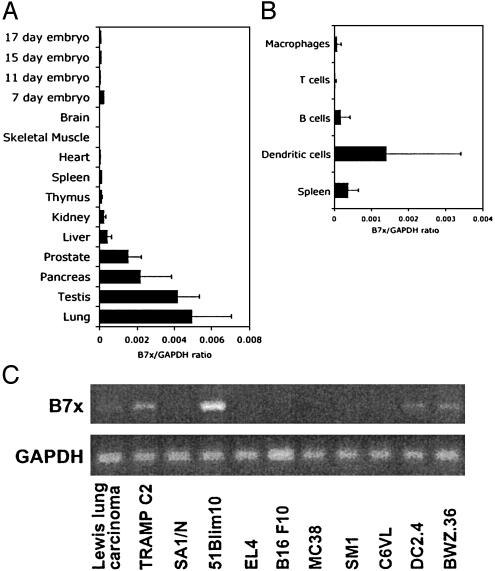
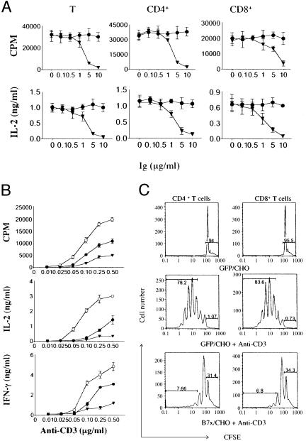
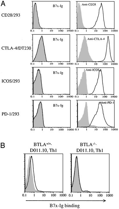
B7 family proteins provide costimulatory signals that regulate T cell responses. Here we report the third set of B7 family-related T cell inhibitory molecules with the identification of a homolog of the B7 family, B7x. It is expressed in immune cells, nonlymphoid tissues, and some tumor cell lines. B7x inhibits cell-cycle progression, proliferation, and cytokine production of both CD4+ and CD8+ T cells. B7x binds a receptor that is expressed on activated, but not resting T cells that is distinct from known CD28 family members. Its receptor may be a recently identified inhibitory molecule, B and T lymphocyte attenuator. These studies identify a costimulatory pathway that may have a unique function in downregulation of tissue-specific autoimmunity and antitumor responses.


Figures

Fig. 1.
Fig. 1.
B7x is a member of the B7 family. (A) Comparison of human B7x with mouse B7x. Predicted signal peptide, Ig V-like and C-like domains, and the hydrophobic/transmembrane region are indicated. Identical amino acids are highlighted in black, and similar residues are shaded in gray. The potential N-glycosylation sites are indicated by arrows. (B) Phylogenetic tree of mouse and human B7 family generated by paup* version 4.0b10. Numbers show the percentage of bootstrap support for each clade.
Fig. 2.
Fig. 2.
B7x is widely expressed in multiple tissues and tumor lines. (A) Real-time PCR was performed on cDNA from multiple mouse tissues. cDNA from the CLONTECH mouse multiple-tissue cDNA panel I was used as well as cDNA made from tissues dissected out of two C57/BL6 mice. The error bars represent the SD among the different mouse cDNA samples. (B) Real-time PCR was performed on CD11c+ dendritic cells, B cells, and T cells that were purified from the spleen and compared to the whole spleen. Thioglycolate-induced macrophages were purified by overnight adherence and removal of nonadherent cells. The results shown are the average and SD among 4-10 individual mouse samples. (C) RT-PCR analysis of B7x expression in mouse tumor cell lines.
Fig. 3.
Fig. 3.
B7x inhibits T cell activation. (A) Purified T cells and CD4+/CD8+ subsets from BALB/c mice were stimulated with plate-bound anti-CD3 (0.25 μg/ml for CD4+ and total T cells, 2 μg/ml for CD8+ T cells) and plate-bound B7x-Ig (▾) or control Ig (•). Aliquots of supernatants were collected 48 h after initiation of cultures, cytokines were measured by ELISA, and proliferation was measured after 72 h by thymidine incorporation. Error bars indicate SD of triplicate cultures. These data are representative of three independent experiments. (B) Purified T cells were stimulated with plate-bound anti-CD3 and CHO transfectants expressing GFP (•), B7-2 (○), or B7x (▾). Cytokines and proliferation were measured as above. These data are representative of five independent experiments. (C) T cells were labeled with CFSE and stimulated with or without plate-bound anti-CD3 and CHO transfectants expressing GFP or B7x. On day 4, cells were harvested, stained with PE-anti-CD4 or PE-anti-CD8, and analyzed by flow cytometry. Percentages refer to fraction of cells in the nondividing peak or cells that have divided more than twice. These data are representative of three independent experiments.
Fig. 4.
Fig. 4.
B7x has a putative counterreceptor on activated T cells distinct from CD28, CTLA-4, ICOS, and PD-1. (A) The 293 transfectants expressing CD28, ICOS, and PD-l or the DT230 transfectant expressing cell surface CTLA-4 were stained with B7x-Ig fusion protein (open histograms) or mouse IgG1 (shaded histograms) as a control, and were then stained with a PE-conjugate anti-mouse-IgG. All transfectants were also stained with specific mAbs (open histograms) or control Abs (shaded histograms). (B) DO11.10 T cell antigen receptor transgenic Th1 cells from WT or BTLA-/- mice were incubated with irradiated BALB/c spleen cells in the presence of 5 μg/ml OVA323-339 peptide for 4 days. Cells were then incubated with B7x-Ig (open histograms) or human IgG1 (shaded histograms) as a control and stained with a PE-conjugated anti-human-IgG.

References

    1. Egen, J. G., Kuhns, M. S. & Allison, J. P. (2002) Nat. Immunol. 3, 611-618. - PubMed
    1. Sharpe, A. H. & Freeman, G. J. (2002) Nat. Rev. Immunol. 2, 116-126. - PubMed
    1. Abbas, A. K. & Sharpe, A. H. (1999) Nat. Med. 5, 1345-1346. - PubMed
    1. Coyle, A. J. & Gutierrez-Ramos, J. C. (2001) Nat. Immunol. 2, 203-209. - PubMed
    1. Carreno, B. M. & Collins, M. (2002) Annu. Rev. Immunol. 20, 29-53. - PubMed

Publication types

MeSH terms