. 2003 Aug;139(8):1505-15.
doi: 10.1038/sj.bjp.0705391.

Cyclooxygenase-1 and cyclooxygenase-2 in the mouse ductus arteriosus: individual activity and functional coupling with nitric oxide synthase

Affiliations

Cyclooxygenase-1 and cyclooxygenase-2 in the mouse ductus arteriosus: individual activity and functional coupling with nitric oxide synthase

B Baragatti et al. Br J Pharmacol. 2003 Aug.

Abstract

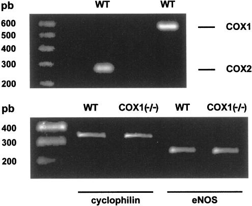
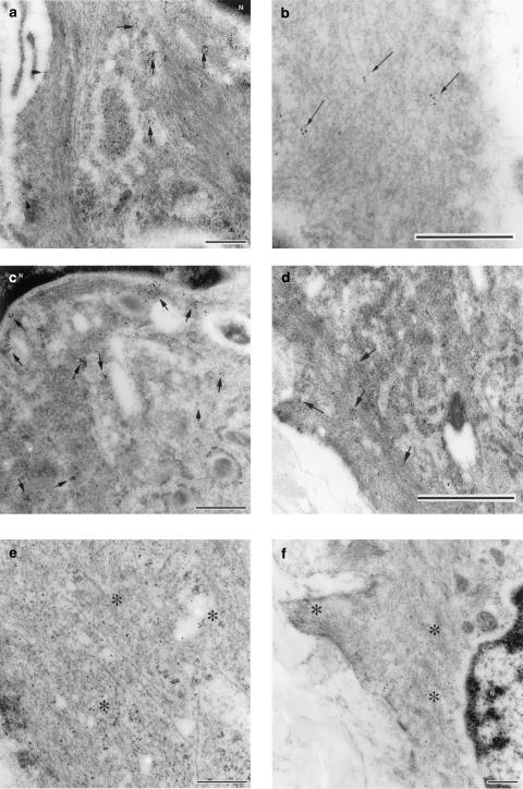
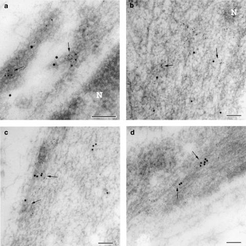
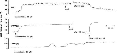
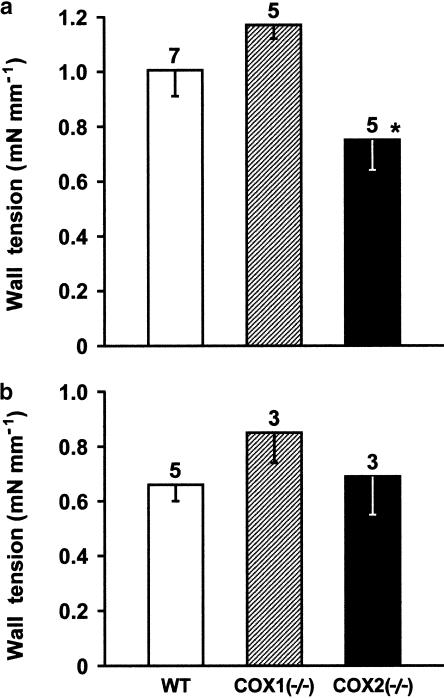
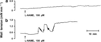
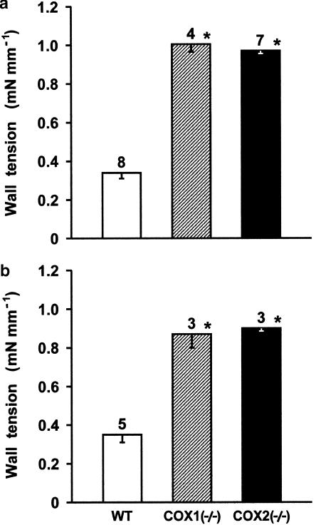
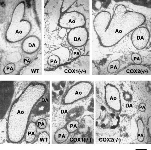
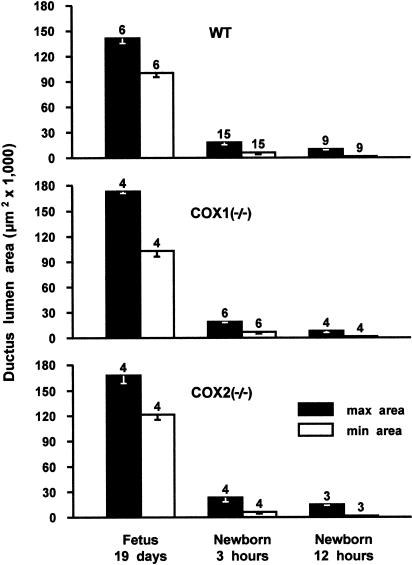
1. Prenatal patency of the ductus arteriosus is maintained by prostaglandin (PG) E(2), conceivably in concert with nitric oxide (NO). Local PGE(2) formation is sustained by cyclooxygenase-1 (COX1) and cyclooxygenase-2 (COX2), a possible exception being the mouse in which COX1, or both COXs, are reportedly absent. Here, we have examined the occurrence of functional COX isoforms in the near-term mouse ductus and the possibility of COX deletion causing NO upregulation. 2. COX1 and COX2 were detected in smooth muscle cells by immunogold electronmicroscopy, both being located primarily in the perinuclear region. Cytosolic and microsomal PGE synthases (cPGES and mPGES) were also found, but they occurred diffusely across the cytosol. COX1 and, far more frequently, COX2 were colocalised with mPGES, while neither COX appeared to be colocalized with cPGES. 3. The isolated ductus from wild-type and COX1-/- mice contracted promptly to indomethacin (2.8 micro M). Conversely, the contraction of COX2-/- ductus to the same inhibitor started only after a delay and was slower. 4. N(G)-nitro-L-arginine methyl ester (L-NAME, 100 micro M) weakly contracted the isolated wild-type ductus. Its effect, however, increased three- to four-fold after deleting either COX, hence equalling that of indomethacin. 5. In vivo, the ductus was patent in all mice foetuses, whether wild-type or COX-deleted. Likewise, no genotype-related difference was noted in its postnatal closure. 6. We conclude that the mouse ductus has a complete system for PGE(2) synthesis comprising both COX1 and COX2. The two enzymes respond differently to indomethacin but, nevertheless, deletion of either one results in NO upregulation. PGE(2) and NO can function synergistically in keeping the ductus patent.


Figures

Figure 1
Figure 1
Gel electrophoresis of RT–PCR-amplified COX (upper panel) and eNOS (lower panel) isoforms of mRNA in the foetal ductus arteriosus from wild-type (WT) mice. Note that the level of eNOS transcript was normalised to that of cyclophilin and did not change upon COX1 deletion (lower panel). COX2−/− animals were not used since they behave as the COX1−/− animals in showing an enhanced NOS-based relaxing activity (see text and Figure 8).
Figure 2
Figure 2
Transmission immunoelectron microscopy from the muscle layer of the foetal mouse ductus arteriosus at term (n=4). (a) COX1 and COX2 immunogold labelling of the whole cytoplasm (cyt) and its perinuclear and peripheral components. Note that gold particle size was different in the two analyses (whole cytoplasm: 5 nm; perinuclear/peripheral cytoplasm: 10 nm). (b) Immunogold labelling for microsomal and cytosolic PGE synthase (mPGES and cPGES). *vs cytoplasm perinuclear, P<0.05 (Student's t-test); vs COX1, whole cytoplasm, P<0.01 (Student's t-test).
Figure 3
Figure 3
Transmission electron micrograph of ultrathin cryosections from the muscle layer of the foetal mouse ductus arteriosus. (a) Immunogold labelling for COX1; note the gold particles being clustered in the cytoplasm (indicated with arrows) which surrounds the nucleus (N) and in the plasma membrane (arrowheads). (b) Immunogold labelling for COX1 in the peripheral cytoplasm; label (arrows) is not as dense as that seen in the perinuclear cytoplasm. (c) Immunogold labelling for COX2 in the perinuclear cytoplasm; note clusters of label in proximity of the nucleus (N). (d) Immunogold labelling for COX2; note few clusters of label (arrows) in the peripheral cytoplasm and along the plasma membrane. (e) Immunogold labelling for cytosolic PGE synthase; label was scattered throughout the cytoplasm (asterisks). (f) Immunogold labelling for microsomal PGE synthase; label was found diffusely throughout the perinuclear (N) and peripheral cytoplasm (asterisks). Bar represents 0.25 μm (panels a, c, e, f) or 0.5 μm (panels b and d).
Figure 4
Figure 4
Transmission electron micrograph of ultrathin cryosections from the muscle layer of the foetal mouse ductus arteriosus. (a) Dual immunogold labelling for COX1 (particle, 10 nm) and microsomal PGE synthase (particle, 5 nm); note colocalisation of the two labels (arrows) in vesicle-like structures close to the nucleus (N). (b) Dual immunogold labelling for COX2 (particle, 10 nm) and microsomal PGE synthase (particle, 5 nm); colocalisation of the two labels (arrows) in proximity of the nucleus (N). (c) Dual immunogold labelling for COX1 (particle, 10 nm) and microsomal PGE synthase (particle, 5 nm) in the peripheral cytoplasm; colocalisation of the two labels in vesicle-like structures (arrows). (d) Dual immunogold labelling for COX2 (particle, 10 nm) and microsomal PGE synthase (particle, 5 nm) in the peripheral cytoplasm; colocalisation of the two labels (arrows). Note that in both perinuclear and peripheral cytoplasm, COX2 colocalised more frequently than COX1 with microsomal PGE synthase. Bar represents 0.1 μm.
Figure 5
Figure 5
Representative responses to indomethacin in the wild-type (WT) vs COX-deleted foetal mouse ductus arteriosus. Contraction to the TXA2 analogue, ONO-11113, given as a reference. Length of the vessel is 540, 444 and 481 μm (short side, see Methods) for wild-type, COX1−/− and COX2−/−, respectively.
Figure 6
Figure 6
Comparison of contractile responses to indomethacin (2.8 μM) in the isolated ductus arteriosus from wild-type (WT) vs COX-deleted mouse foetus. (a) 2.5% O2; (b) 12.5% O2. Wall tension (mN mm−1) prior to treatment, respectively, at 2.5 and 12.5% O2, was as follows: WT, 0.36±0.04 and 0.36±0.15; COX1−/−, 0.30±0.03 and 0.29±0.13; COX2−/−, 0.25±0.08 and 0.36±0.03. For each group, the number of experiments is given above each column, and no significant difference between wild-type and COX-deleted preparations is reached at either O2 tension. A significant difference in the panel (a) relative to COX1−/− is indicated with *P<0.05 (ANOVA). Note that in the case of wild-type and COX1−/− preparations, responses are slightly lower at 12.5 than 2.5% O2 (P<0.05).
Figure 7
Figure 7
Representative response to L-NAME in the wild-type vs COX-deleted foetal mouse ductus arteriosus. (a) wild-type; (b) COX1−/− mutant. Note that response pattern is similar in COX1−/− and COX2−/−. Length of the vessel is 540 and 548 μm (short side, see Methods) for wild-type and COX1−/−, respectively.
Figure 8
Figure 8
Comparison of contractile responses to L-NAME (100 μM) in the isolated ductus arteriosus from wild-type (WT) vs COX-deleted mouse foetus. (a) 2.5% O2; (b) 12.5% O2. Wall tension (mN mm−1) prior to treatment, respectively, at 2.5 and 12.5% O2, was as follows: WT, 0.07±0.03 and 0.37±0.04; COX1−/−, 0.24±0.1 and 0.14±0.08; COX2−/−, 0.08±0.05 and 0.2±0.12. For each group, the number of experiments is given above the columns, and a significant difference (P<0.01) between wild-type and COX-deleted preparations is indicated with an asterisk.
Figure 9
Figure 9
Cross-section of the ductus arteriosus and the large blood vessels in the wild-type and COX-deleted frozen mice. (upper panel) Foetus at 19 days gestation; (lower panel) newborn at 3 h. WT, wild-type; DA, ductus arteriosus; Ao, aorta; PA, pulmonary artery. Scale bar, 250 μm.
Figure 10
Figure 10
Time course of ductus closure in wild-type (WT) and COX-deleted mice. For each group, the number of animals is given above the columns.

Similar articles

Cited by

References

    1. BALLOU L.R., BOTTING R.M., GOORHA S., ZHANG J., VANE J.R. Nociception in cyclooxygenase isozyme-deficient mice. Proc. Natl. Acad. Sci. U.S.A. 2000;97:10272–10276. - PMC - PubMed
    1. BOUAYAD A., KAJINO H., WALEH N., FOURON J.-C., ANDELFINGER G., VARMA D.R., SKOLL A., VAZQUEZ A., GOBEIL F., JR, CLYMAN R.I., CHEMTOB S. Characterisation of PGE receptors in fetal and newborn lamb ductus arteriosus. Am. J. Physiol. 2001;280:H2342–H2349. - PubMed
    1. CHALLIS J.R.G., DILLEY S.R., ROBINSON J.S., THORBURN G.D. Prostaglandins in the circulation of the fetal lamb. Prostaglandins. 1976;11:1041–1052. - PubMed
    1. CHOMCZYNSKY P., SACCHI N. Single-step method of RNA isolation by acid guanidinium thyocyanate–phenol–chloroform extraction. Anal. Biochem. 1987;162:156–159. - PubMed
    1. CLYMAN R.I. Ductus arteriosus: current theories of prenatal and postnatal regulation. Semin. Perinatol. 1987;11:64–71. - PubMed

Publication types

LinkOut - more resources