. 2003 Sep;62(9):851-8.
doi: 10.1136/ard.62.9.851.

Perturbations of peripheral B lymphocyte homoeostasis in children with systemic lupus erythematosus

Affiliations

Perturbations of peripheral B lymphocyte homoeostasis in children with systemic lupus erythematosus

M Odendahl et al. Ann Rheum Dis. 2003 Sep.

Abstract

Objective: To investigate the distribution of peripheral B cell subpopulations of children with active and inactive systemic lupus erythematosus (SLE) compared with healthy controls.

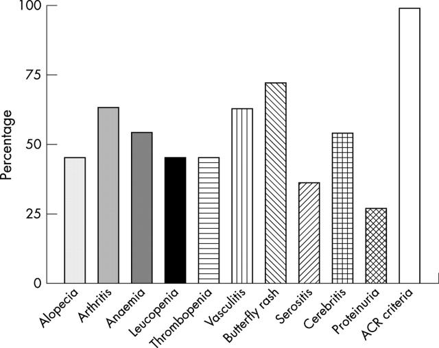
Methods: Peripheral B cell subpopulations of 11 children with SLE (6 with active and 5 with inactive disease) and 14 age matched normal healthy children were analysed. Active disease was diagnosed in children with a flare of SLE, who received treatment by i.v. cyclophosphamide or i.v. methylprednisolone pulse to control the disease. Additionally, the peripheral B cells of the children with SLE were compared with those of 13 consecutive patients with adult onset SLE.

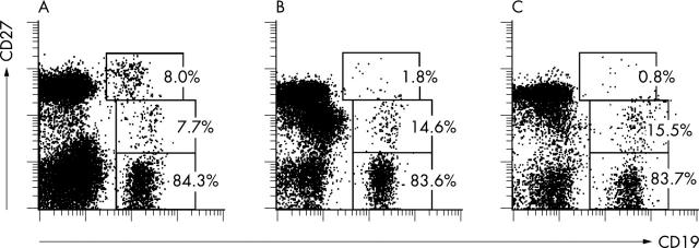
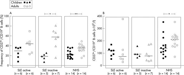
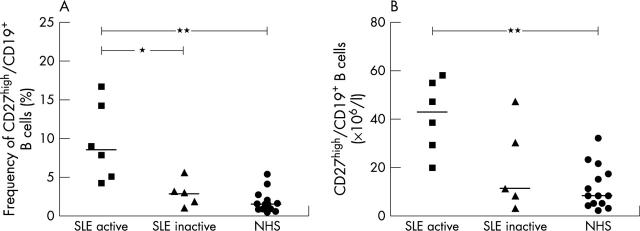
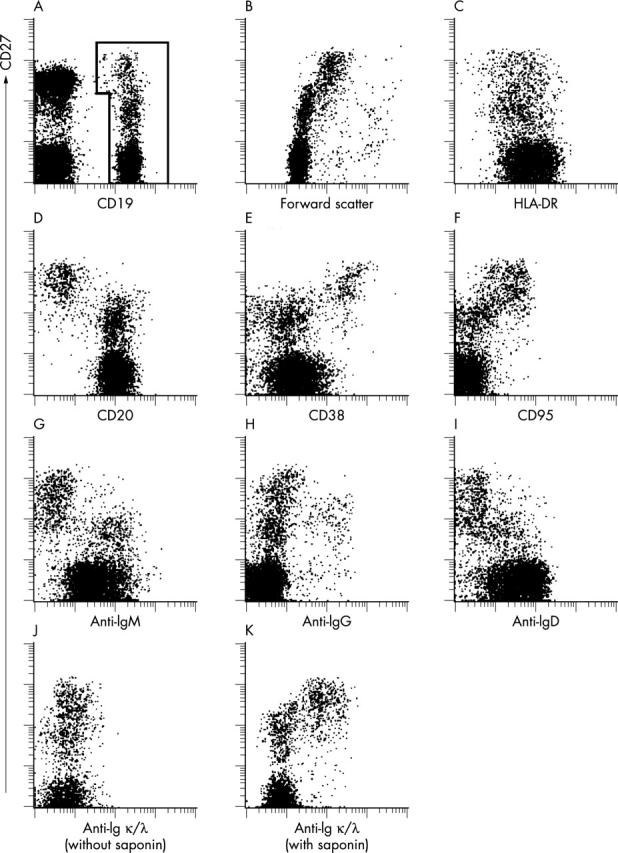
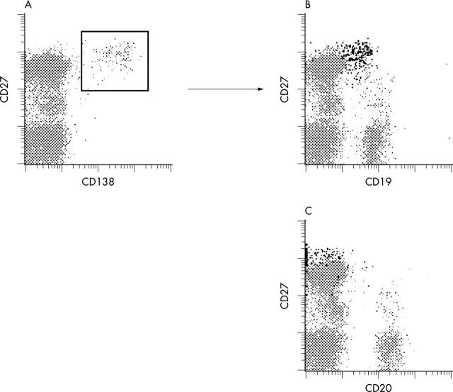
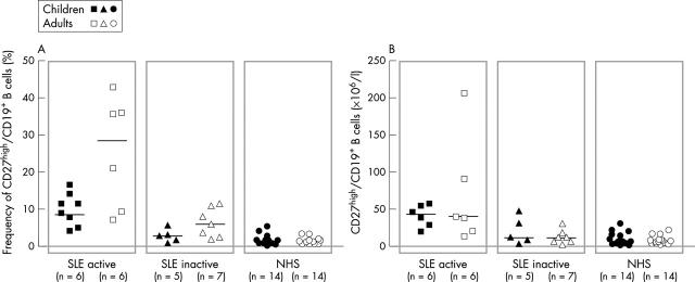
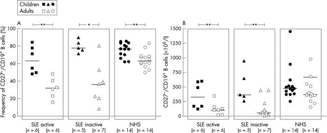
Results: No major difference was found in the frequency and total number of CD27(-)/CD19(+) naïve B cells and CD27(+)/CD19(+) memory B cells between patients with active and inactive lupus and healthy controls, but there was a significant increase in CD27(high) expressing plasma blasts in patients with active SLE. These cells coexpress CD38(+), HLA-DR(dim), surface Ig(low) and lack the expression of CD20 but are clearly positive for intracellular Ig, indicative of early plasma cells. Most CD138(+) cells coexpress CD27(high)/CD19(+). The enhanced frequency of peripheral plasma blasts in children with active SLE is consistent with previous findings in adult patients with SLE, whereas a relative predominance of CD27(+) memory B cells was only identified in the adult patients.

Conclusions: Profound abnormalities in the distribution of B cell compartments are more pronounced in older patients with SLE, but an enhanced frequency and cell number of peripheral plasma blasts is characteristic of both diseases during active stages. Thus detection of CD27(high) plasma blasts significantly correlates with active lupus in both children and adults.


Figures

Figure 1
Figure 1
Frequency of clinical manifestations of the children with SLE analysed in the current study. All analysed children showed at least four different clinical manifestations classifying them according to the 1982 revised ACR criteria.
Figure 2
Figure 2
Expression of CD27 on CD19+ peripheral B cells from children with SLE and from a normal healthy donor. Viable PBMC were gated for analysis according to light scatter and exclusion of propidium iodide. Staining with CD19 bio/SA-PE versus CD27 Cy5 is shown for (A) a child with active SLE (donor No 6); (B) a child with inactive SLE (donor No 3); and (C) a healthy child recruited outside the hospital (donor No 12). Gates for the statistical evaluation of the frequency of CD27-, CD27+, and CD27high among CD19+ peripheral B cells are indicated. The respective frequencies are indicated.
Figure 7
Figure 7
Comparison of the (A) frequency and (B) total numbers of CD27+ peripheral memory B cells in children and adults from a previous study of patients with active and inactive SLE as well as in their respective healthy controls. The frequencies were determined by cytometric analysis as shown in fig 2. The median values are indicated. *p<0.05; **p<0.01.
Figure 3
Figure 3
Frequency (A) and total number (B) of CD27high peripheral plasma cells in patients with active and inactive SLE and in a control group of healthy subjects. The frequencies were determined by cytometric analysis as shown in fig 2. The median value is indicated. *p<0.05; **p<0.01.
Figure 4
Figure 4
Cytometric characterisation of peripheral CD19+ B cells in a child with SLE (donor No 2). (A) Viable peripheral mononuclear cells of a patient with SLE, gated according to scatter and propidium iodide exclusion, were tested for CD19 and CD27 expression. CD19+ B cells were gated for further analysis as indicated. (B) Staining of CD27 versus forward light scatter to analyse differences in cell size. (C-I) CD19+ B cells, as gated in (A), were counterstained for HLA-DR, CD20, CD38, CD95, IgM, IgG, or IgD, respectively. Formaldehyde fixed cells were gated according to scatter and CD19-PE staining and counterstained for CD27 and for Igκ and Igλ light chains, without (J) or with (K) permeabilisation of the cell membrane by the use of saponin.
Figure 5
Figure 5
Cytometric characterisation of peripheral CD138+ cells in a child with a lupus flare (donor No 6). (A) Viable peripheral mononuclear cells of a patient with a lupus flare were gated according to scatter and propidium iodide exclusion and stained for CD138-PE, CD27-Cy5, and either CD19-FITC or CD20-FITC, respectively. Coexpression of CD27 and CD19 (B) or CD20 (C), respectively, on CD138+ cells, as gated in (A), is shown in a two colour flow cytometric analysis. CD138+ coexpressing cells in (B) and (C) are depicted in black.
Figure 8
Figure 8
Comparison of the (A) frequency and (B) total numbers of CD27high peripheral plasma cells in children and adults from a previous study of patients with active and inactive SLE as well as in their respective healthy controls. The frequencies were determined by cytometric analysis as shown in fig 2. The median values are indicated. No significant difference was found between the children and adults with SLE.
Figure 6
Figure 6
Comparison of the (A) frequency and (B) total numbers of CD27- peripheral naïve B cells in children and adults from a previous study of patients with active and inactive SLE as well as their respective healthy controls. The frequencies were determined by cytometric analysis as shown in fig 2. The median values are indicated. *p<0.05; **p<0.01.

References

    1. J Immunol. 1998 Dec 15;161(12):6496-502 - PubMed
    1. J Exp Med. 1998 Nov 2;188(9):1691-703 - PubMed
    1. Immunol Rev. 1999 Jun;169:107-21 - PubMed
    1. Blood. 1999 Nov 15;94(10):3551-8 - PubMed
    1. J Immunol. 2000 Nov 15;165(10):5970-9 - PubMed

Publication types

Substances