Comparative Study
. 2003 Aug 15;17(16):2060-72.
doi: 10.1101/gad.1103303.

Structure of HoxA9 and Pbx1 bound to DNA: Hox hexapeptide and DNA recognition anterior to posterior

Affiliations
Comparative Study

Structure of HoxA9 and Pbx1 bound to DNA: Hox hexapeptide and DNA recognition anterior to posterior

Nicole A LaRonde-LeBlanc et al. Genes Dev. .

Abstract

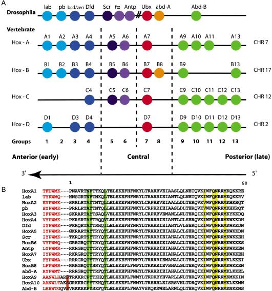
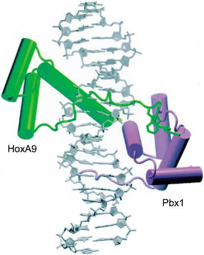
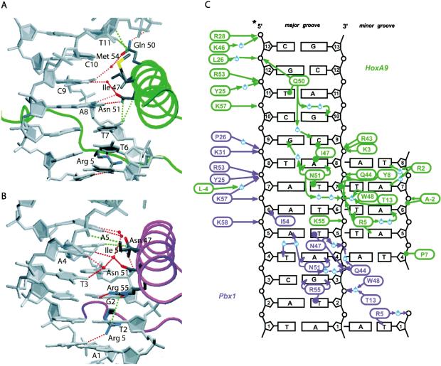
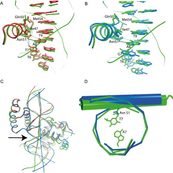
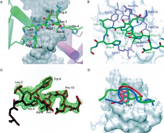
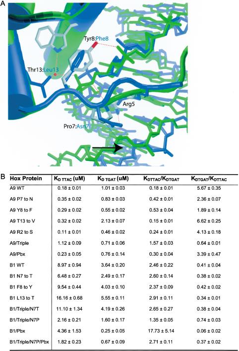
The HOX/HOM superfamily of homeodomain proteins controls cell fate and segmental embryonic patterning by a mechanism that is conserved in all metazoans. The linear arrangement of the Hox genes on the chromosome correlates with the spatial distribution of HOX protein expression along the anterior-posterior axis of the embryo. Most HOX proteins bind DNA cooperatively with members of the PBC family of TALE-type homeodomain proteins, which includes human Pbx1. Cooperative DNA binding between HOX and PBC proteins requires a residue N-terminal to the HOX homeodomain termed the hexapeptide, which differs significantly in sequence between anterior- and posterior-regulating HOX proteins. We report here the 1.9-A-resolution structure of a posterior HOX protein, HoxA9, complexed with Pbx1 and DNA, which reveals that the posterior Hox hexapeptide adopts an altered conformation as compared with that seen in previously determined anterior HOX/PBC structures. The additional nonspecific interactions and altered DNA conformation in this structure account for the stronger DNA-binding affinity and altered specificity observed for posterior HOX proteins when compared with anterior HOX proteins. DNA-binding studies of wild-type and mutant HoxA9 and HoxB1 show residues in the N-terminal arm of the homeodomains are critical for proper DNA sequence recognition despite lack of direct contact by these residues to the DNA bases. These results help shed light on the mechanism of transcriptional regulation by HOX proteins and show how DNA-binding proteins may use indirect contacts to determine sequence specificity.


Figures

Figure 1.
Figure 1.
(A) Arrangement of the Hox genes of the Drosophila and vertebrate clusters. The arrangement 5′ to 3′ is conserved across these species. Members of the same paralog group are colored identically. Abd-B-like Hox genes are shown in green. (B) Alignment of the homeodomain sequences in groups 1–10 of Drosophila and vertebrates shows extensive sequence conservation. Broken lines indicate where linkers of variable lengths would be. The three-amino-acid linkers of the Abd-B-like Hox proteins are shown with the conserved Ser –1 highlighted in orange. The hexapeptides are shown in red text, and the DNA recognition residues are highlighted in yellow. Positions 7, 8, and 13 of the homeodomains are highlighted in green.
Figure 2.
Figure 2.
The HoxA9–Pbx1–DNA complex. HoxA9 is shown in green, Pbx1 in purple. The linker and the hexapeptide of HoxA9 spans the minor groove (front), and the conserved tryptophan is inserted in the Pbx1 binding pocket. This figure and subsequent structure graphics were made using VMD.
Figure 4.
Figure 4.
DNA contacts made by HoxA9 (A) and Pbx1 (B). Minor groove interactions are made by Arg 5 in the back for both homeodomains. Hydrophobic interactions (green) and hydrogen bonds (red) are shown as broken lines. (C) Schematic of HoxA9 (green) and Pbx1 (purple) DNA interactions. Hydrogen bonds are indicated with arrows, and van der Waals interactions are shown as lines terminating in circles. Minor groove interactions are on the left side of the figure. The HoxA9/Pbx1 site is 5′-A1T2G3A4T5T6T7A8C9G10A11C12-3′.
Figure 5.
Figure 5.
Alignment of the third helix of HoxA9 (green) with Ubx (A; red) and HoxB1 (B; blue). The interaction of Asn 51 with T7 is conserved in Ubx, but may not be made in HoxB1. (C) Alignment of the full homeodomain of Ubx (red) and HoxB1 (blue) with HoxA9 (green) shows that the DNA in both the HoxA9 and Ubx complexes contains a bend (arrow) relative to HoxB1. (D) Alignment of the DNA in the region of the Hox binding sites of HoxA9 (green) and HoxB1 (blue) shows a shift of the HoxA9 recognition helix and Asn 51 into the major groove relative to HoxB1.
Figure 3.
Figure 3.
The linker and the hexapeptide of HoxA9. (A) Detailed view of the N-terminal arm and the linker region of the HoxA9 homeodomain spanning the minor groove. The linker region is composed of Ala –3, Arg –2, and Ser –1. Arg 2 makes water-mediated contacts in the minor groove, and Lys 4 and Ala –3 contact the sugar-phosphate backbone. (B) The HoxA9 hexapeptide (green) in the Pbx1 hexapeptide-binding pocket (purple residues and white surface). Hydrophobic interactions and hydrogen bonds between HoxA9 and Pbx1 are shown in gray and blue, respectively. The TALE insertion in the Pbx1 homeodomain is composed of Leu 23a, Ser 23b, and Asn 23c. (C) Simulated annealing omit map contoured at 3 σ for residues –12 to –4 of HoxA9. The hexapeptide residues as well as additional residues in the N terminus are shown. (D) Comparison of the hexapeptide backbone conformations of HoxA9 (green), HoxB1 (blue), and Ubx (red). The PBC homeodomains of the HoxB1 and Ubx complexes were aligned with the Pbx1 homeodomain (white) of the HoxA9 complex to overlay the peptides. The conserved tryptophan is labeled as Trp –6.
Figure 6.
Figure 6.
DNA-binding studies of HoxA9 and HoxB1. (A) Residues 7, 8, and 13 of HoxA9 contact the sugar-phosphate backbone and allow Arg 5 to make contact across the minor groove in HoxA9, whereas in HoxB1 no such contacts are observed. (B) Table of equilibrium dissociation constants measured by gel retardation assays using wild-type and mutant HoxA9 and HoxB1 on DNA containing the consensus binding sites for HoxA9 (TTTAC) and HoxB1 (TTGAT).

References

    1. Berry, M. and Gehring, W. 2000. Phosphorylation status of the SCR homeodomain determines its functional activity: Essential role for protein phosphatase 2A,B′. EMBO J. 19: 2946–2957. - PMC - PubMed
    1. Billeter, M., Qian, Y.Q., Otting, G., Muller, M., Gehring, W., and Wuthrich, K. 1993. Determination of the nuclear magnetic resonance solution structure of an Antennapedia homeodomain–DNA complex. J. Mol. Biol. 234: 1084–1093. - PubMed
    1. Brünger, A.T., Adams, P.D., Clore, G.M., DeLano, W.L., Gros, P., Grosse-Kunstleve, R.W., Jiang, J.S., Kuszewski, J., Nilges, M., Pannu, N.S., et al. 1998. Crystallography & NMR system: A new software suite for macromolecular structure determination. Acta Crystallogr. D Biol. Crystallogr. 54 (Pt 5): 905–921. - PubMed
    1. Bürglin, T.R. 1997. Analysis of TALE superclass homeobox genes (MEIS, PBC, KNOX, Iroquois, TGIF) reveals a novel domain conserved between plants and animals. Nucleic Acids Res. 25: 4173–4180. - PMC - PubMed
    1. Chan, S.K., Jaffe, L., Capovilla, M., Botas, J., and Mann, R.S. 1994. The DNA binding specificity of Ultrabithorax is modulated by cooperative interactions with extradenticle, another homeoprotein. Cell 78: 603–615. - PubMed

Publication types

MeSH terms

Associated data

LinkOut - more resources