

Membrane localization of motility, signaling, and polyketide synthetase proteins in Myxococcus xanthus
- PMID: 12923079
- PMCID: PMC181019
- DOI: 10.1128/JB.185.17.5066-5075.2003
Membrane localization of motility, signaling, and polyketide synthetase proteins in Myxococcus xanthus
Abstract
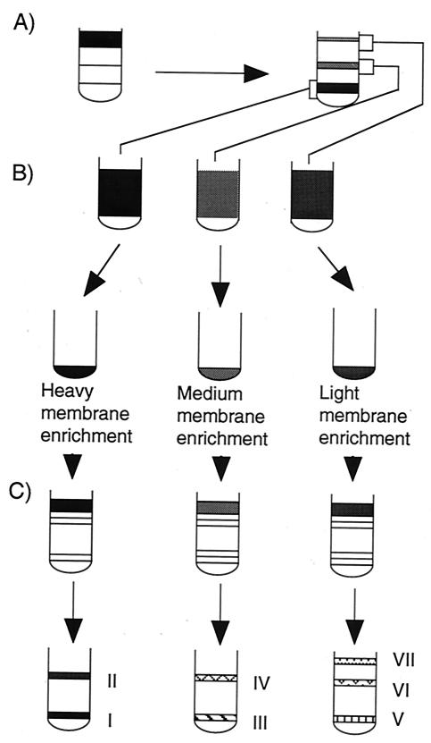
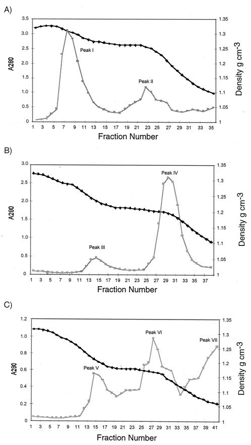
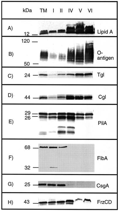
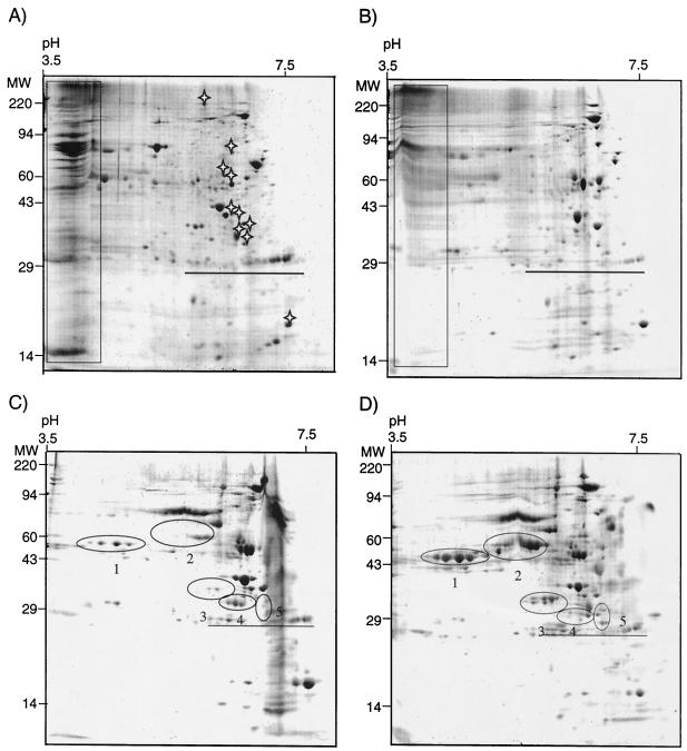
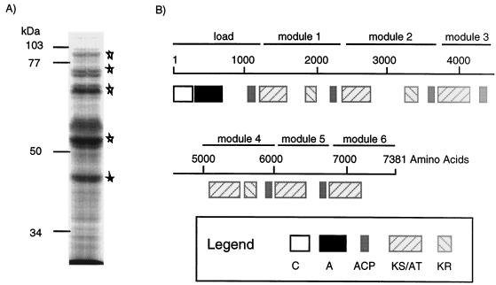
Myxococcus xanthus cells coordinate cellular motility, biofilm formation, and development through the use of cell signaling pathways. In an effort to understand the mechanisms underlying these processes, the inner membrane (IM) and outer membrane (OM) of strain DK1622 were fractionated to examine protein localization. Membranes were enriched from spheroplasts of vegetative cells and then separated into three peaks on a three-step sucrose gradient. The high-density fraction corresponded to the putative IM, the medium-density fraction corresponded to a putative hybrid membrane (HM), and the low-density fraction corresponded to the putative OM. Each fraction was subjected to further separation on discontinuous sucrose gradients, which resulted in discrete protein peaks for each major fraction. The purity and origin of each peak were assessed by using succinate dehydrogenase (SDH) activity as the IM marker and reactivities to lipopolysaccharide core and O-antigen monoclonal antibodies as the OM markers. As previously reported, the OM markers localized to the low-density membrane fractions, while SDH localized to high-density fractions. Immunoblotting was used to localize important motility and signaling proteins within the protein peaks. CsgA, the C-signal-producing protein, and FibA, a fibril-associated protease, were localized in the IM (density, 1.17 to 1.24 g cm(-3)). Tgl and Cgl lipoproteins were localized in the OM, which contained areas of high buoyant density (1.21 to 1.24 g cm(-3)) and low buoyant density (1.169 to 1.171 g cm(-3)). FrzCD, a methyl-accepting chemotaxis protein, was predominantly located in the IM, although smaller amounts were found in the OM. The HM peaks showed twofold enrichment for the type IV pilin protein PilA, suggesting that this fraction contained cell poles. Two-dimensional polyacrylamide gel electrophoresis revealed the presence of proteins that were unique to the IM and OM. Characterization of proteins in an unusually low-density membrane peak (1.072 to 1.094 g cm(-3)) showed the presence of Ta-1 polyketide synthetase, which synthesizes the antibiotic myxovirescin A.
Figures
Similar articles
-
Myxobacterial tools for social interactions.Res Microbiol. 2012 Nov-Dec;163(9-10):579-91. doi: 10.1016/j.resmic.2012.10.022. Epub 2012 Nov 2. Res Microbiol. 2012. PMID: 23123306 Free PMC article. Review.
-
Identification and localization of Myxococcus xanthus porins and lipoproteins.PLoS One. 2011;6(11):e27475. doi: 10.1371/journal.pone.0027475. Epub 2011 Nov 22. PLoS One. 2011. PMID: 22132103 Free PMC article.
-
Predominant outer membrane antigens of Bartonella henselae.Infect Immun. 2004 Jun;72(6):3097-105. doi: 10.1128/IAI.72.6.3097-3105.2004. Infect Immun. 2004. PMID: 15155610 Free PMC article.
-
Identification and localization of the Tgl protein, which is required for Myxococcus xanthus social motility.J Bacteriol. 1997 Jul;179(13):4372-81. doi: 10.1128/jb.179.13.4372-4381.1997. J Bacteriol. 1997. PMID: 9209056 Free PMC article.
-
Cell polarity, intercellular signalling and morphogenetic cell movements in Myxococcus xanthus.Curr Opin Microbiol. 2004 Dec;7(6):587-93. doi: 10.1016/j.mib.2004.10.004. Curr Opin Microbiol. 2004. PMID: 15556030 Review.
Cited by
-
The biosynthetic genes encoding for the production of the dynemicin enediyne core in Micromonospora chersina ATCC53710.FEMS Microbiol Lett. 2008 May;282(1):105-14. doi: 10.1111/j.1574-6968.2008.01112.x. Epub 2008 Mar 5. FEMS Microbiol Lett. 2008. PMID: 18328078 Free PMC article.
-
A singular enzymatic megacomplex from Bacillus subtilis.Proc Natl Acad Sci U S A. 2007 Jan 2;104(1):305-10. doi: 10.1073/pnas.0609073103. Epub 2006 Dec 26. Proc Natl Acad Sci U S A. 2007. PMID: 17190806 Free PMC article.
-
Myxococcus CsgA, Drosophila Sniffer, and human HSD10 are cardiolipin phospholipases.Genes Dev. 2015 Sep 15;29(18):1903-14. doi: 10.1101/gad.268482.115. Epub 2015 Sep 3. Genes Dev. 2015. PMID: 26338420 Free PMC article.
-
Myxobacterial tools for social interactions.Res Microbiol. 2012 Nov-Dec;163(9-10):579-91. doi: 10.1016/j.resmic.2012.10.022. Epub 2012 Nov 2. Res Microbiol. 2012. PMID: 23123306 Free PMC article. Review.
-
Myxobacteria, polarity, and multicellular morphogenesis.Cold Spring Harb Perspect Biol. 2010 Aug;2(8):a000380. doi: 10.1101/cshperspect.a000380. Epub 2010 Jul 7. Cold Spring Harb Perspect Biol. 2010. PMID: 20610548 Free PMC article. Review.
References
MeSH terms
Substances
LinkOut - more resources
Full Text Sources
Research Materials
