. 2003 Sep;185(17):5109-16.
doi: 10.1128/JB.185.17.5109-5116.2003.

Regulation of expression of scaffoldin-related genes in Clostridium thermocellum

Affiliations

Regulation of expression of scaffoldin-related genes in Clostridium thermocellum

Tali W Dror et al. J Bacteriol. 2003 Sep.

Abstract

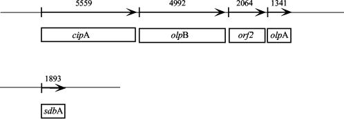
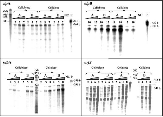
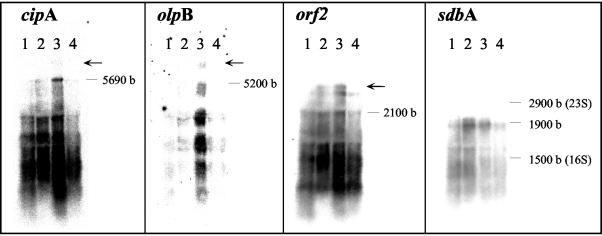
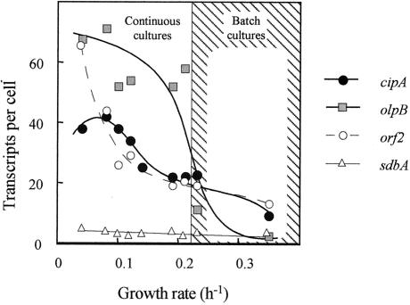
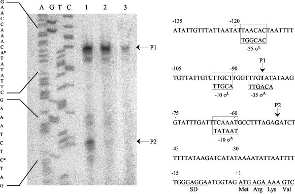
Clostridium thermocellum produces an extracellular multienzyme complex, termed the cellulosome, that allows efficient solubilization of crystalline cellulose. The complex is organized around a large noncatalytic protein subunit, termed CipA or scaffoldin, and is found either free in the supernatant or cell bound. The binding of the complex to the cell is mediated by three cell surface anchoring proteins, OlpB, Orf2p, and SdbA, that interact with the CipA scaffoldin. The transcriptional level of the olpB, orf2, sdbA, and cipA genes was determined quantitatively by RNase protection assays in batch and continuous cultures, under carbon and nitrogen limitation. The mRNA level of olpB, orf2, and cipA varied with growth rate, reaching 40 to 60 transcripts per cell under carbon limitation at a low growth rate of 0.04 h(-1) and 2 to 10 transcripts per cell at a growth rate of 0.35 h(-1) in batch culture. The mRNA level of sdbA was about three transcripts per cell and was not influenced by growth rate. Primer extension analysis revealed two major transcriptional start sites, at -81 and -50 bp, upstream of the translational start site of the cipA gene. The potential promoters exhibited homology to the known sigma factors sigma(A) and sigma(L) (sigma(54)) of Bacillus subtilis. Transcription from the sigma(L)-like promoter was found under all growth conditions, whereas transcription from the sigma(A)-like promoter was significant only under carbon limitation. The overall expression level obtained in the primer extension analysis was in good agreement with the results of the RNase-protection assays.


Figures

FIG. 1.
FIG. 1.
Organization of C. thermocellum scaffoldin-related genes. cipA and the three genes olpB, orf2, and olpA are located in tandem within a gene cluster. sdbA is located on a distinct region of the chromosome. The proteins OlpB, Orf2p, and SdbA serve as anchoring proteins that connect the CipA scaffoldin to the cell surface. In contrast, OlpA selectively binds individual cellulases to the cell surface. The numbers above the gene in the scheme refer to its size (in base pairs).
FIG. 2.
FIG. 2.
Transcript levels of the cipA gene and the genes coding for the cipA-specific anchoring proteins, olpB, orf2, and sdbA. RPAs were performed with the relevant 32P-labeled antisense probe as described earlier (11), using RNA from exponential (A) or stationary (B) phase cultures, grown on either cellobiose or crystalline cellulose. Different amounts (in micrograms) of RNA hybridized with the probes are indicated at the top of each lane. Autoradiographs of the respective RPA products were visualized using a phosphorimager system. Negative control lanes (NC) contained RNA from yeast. Lane M corresponds to markers (in base pairs). The full-length probe is shown in lane P. Values at right indicate the sizes of the undigested full-length antisense probes and the estimated sizes of the protected products in bases (b).
FIG. 3.
FIG. 3.
mRNA expression of cipA and scaffoldin-related genes as determined by Northern blot analysis. Northern blot analysis was performed using total RNA isolated from cultures grown on either cellobiose (lanes 1 and 2) or crystalline cellulose (lanes 3 and 4), during exponential (lanes 1 and 3) and stationary (lanes 2 and 4) phases of growth. The isolated RNA (15 μg) was loaded and separated on agarose gels and then transferred to nitrocellulose membranes. The blot was hybridized subsequently with cipA, olpB, sdbA, and orf2 probes. The sizes of the16S and 23S rRNA and the estimated sizes of the transcripts are indicated in bases (b). Arrows denote larger-than-expected sized transcripts labeled by the designated probe (see text for details).
FIG. 4.
FIG. 4.
Transcript levels of cipA and scaffoldin-related genes as a function of growth rate. The amounts of the respective mRNA were determined by RPA and converted to the number of transcripts per cell based on the average measured amount of total RNA in a single cell (0.18 pg/cell). Values represent the average of several (at least 3) measurements at an accuracy of ± 25%. Continuous cultures were operated under carbon (cellobiose) limitation at dilution rates between 0.04 ≤ μ ≤ 0.21; batch cultures were grown to exponential phase on either cellulose or cellobiose at rates of μ = 0.23 and 0.35 h−1, respectively.
FIG. 5.
FIG. 5.
Mapping of the 5′ terminus of cipA by primer extension analysis. (A) 32P-labeled oligonucleotide was hybridized to mRNA obtained from C. thermocellum grown under the following conditions: continuous culture limited on cellobiose, diluted to a rate of 0.1 h−1 (lane 1); exponential-phase culture grown on Avicel (lane 2); and exponential-phase culture grown on cellobiose (lane 3). Dideoxynucleotide sequence reactions were performed using the same primers employed in the reverse transcriptase reactions. The positions of the transcriptional start points are indicated by asterisks on the inferred non-template-strand sequences. The products of primer extension are indicated by P1 and P2. (B) Sequence data for the regulatory region of cipA. The respective transcriptional start points (P1 and P2) are indicated. The consensus B. subtilis σA and σL promoter sequences are framed at the homologous sites of the C. thermocellum sequence. The proposed Shine-Dalgarno (SD) site and the initiating ATG codon are indicated.

Similar articles

Cited by

References

    1. Bayer, E. A., R. Kenig, and R. Lamed. 1983. Adherence of Clostridium thermocellum to cellulose. J. Bacteriol. 156:818-827. - PMC - PubMed
    1. Bayer, E. A., and R. Lamed. 1986. Ultrastructure of the cell surface cellulosome of Clostridium thermocellum and its interaction with cellulose. J. Bacteriol. 167:828-836. - PMC - PubMed
    1. Bayer, E. A., E. Morag, Y. Shoham, J. Tormo, and R. Lamed. 1996. The cellulosome: a cell-surface organelle for the adhesion to and degradation of cellulose, p. 155-182. In M. Fletcher (ed.), Bacterial adhesion: molecular and ecological diversity. Wiley-Liss, Inc., New York, N.Y.
    1. Bayer, E. A., E. Setter, and R. Lamed. 1985. Organization and distribution of the cellulosome in Clostridium thermocellum. J. Bacteriol. 163:552-559. - PMC - PubMed
    1. Bayer, E. A., L. J. Shimon, Y. Shoham, and R. Lamed. 1998. Cellulosomes: structure and ultrastructure. J. Struct. Biol. 124:221-234. - PubMed

Publication types

MeSH terms

LinkOut - more resources