

Thin pilus PilV adhesins of plasmid R64 recognize specific structures of the lipopolysaccharide molecules of recipient cells
- PMID: 12923092
- PMCID: PMC181018
- DOI: 10.1128/JB.185.17.5192-5199.2003
Thin pilus PilV adhesins of plasmid R64 recognize specific structures of the lipopolysaccharide molecules of recipient cells
Abstract
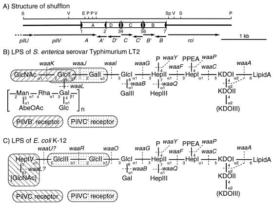
IncI1 plasmid R64 encodes a type IV pilus called a thin pilus, which includes PilV adhesins. Seven different sequences for the C-terminal segments of PilV adhesins can be produced by shufflon DNA rearrangement. The expression of the seven PilV adhesins determines the recipient specificity in liquid matings of plasmid R64. Salmonella enterica serovar Typhimurium LT2 was recognized by the PilVA' and PilVB' adhesins, while Escherichia coli K-12 was recognized by the PilVA', PilVC, and PilVC' adhesins. Lipopolysaccharide (LPS) on the surfaces of recipient cells was previously shown to be the specific receptor for the seven PilV adhesins. To identify the specific receptor structures of LPS for various PilV adhesins, R64 liquid matings were carried out with recipient cells consisting of various S. enterica serovar Typhimurium LT2 and E. coli K-12 waa mutants and their derivatives carrying various waa genes of different origins. From the mating experiments, including inhibition experiments, we propose that the GlcNAc(alpha1-2)Glc and Glc(alpha1-2)Gal structures of the LPS core of S. enterica serovar Typhimurium LT2 function as receptors for the PilVB' and PilVC' adhesins, respectively, while the PilVC' receptor in the wild-type LT2 LPS core may be masked. We further propose that the GlcNAc(beta1-7)Hep and Glc(alpha1-2)Glc structures of the LPS core of E. coli K-12 function as receptors for the PilVC and PilVC' adhesins, respectively.
Figures
Similar articles
-
PilV adhesins of plasmid R64 thin pili specifically bind to the lipopolysaccharides of recipient cells.J Mol Biol. 2004 Oct 22;343(3):615-25. doi: 10.1016/j.jmb.2004.08.059. J Mol Biol. 2004. PMID: 15465049
-
The lipopolysaccharide of recipient cells is a specific receptor for PilV proteins, selected by shufflon DNA rearrangement, in liquid matings with donors bearing the R64 plasmid.Mol Gen Genet. 2000 Feb;263(1):159-64. doi: 10.1007/s004380050043. Mol Gen Genet. 2000. PMID: 10732685
-
DNA rearrangement of the shufflon determines recipient specificity in liquid mating of IncI1 plasmid R64.J Mol Biol. 1994 Oct 14;243(1):6-9. doi: 10.1006/jmbi.1994.1625. J Mol Biol. 1994. PMID: 7932741 Review.
-
Mating variation by DNA inversions of shufflon in plasmid R64.Adv Biophys. 1995;31:181-93. doi: 10.1016/0065-227x(95)99391-2. Adv Biophys. 1995. PMID: 7625273 Review.
-
Structure and function of the shufflon in plasmid R64.Adv Biophys. 2004;38:183-213. Adv Biophys. 2004. PMID: 15493334 Review.
Cited by
-
The Salmonella enterica serovar Typhi type IVB self-association pili are detached from the bacterial cell by the PilV minor pilus proteins.Infect Immun. 2006 Sep;74(9):5414-8. doi: 10.1128/IAI.00172-06. Infect Immun. 2006. PMID: 16926438 Free PMC article.
-
Analysis of the pilU gene for the prepilin peptidase involved in the biogenesis of type IV pili encoded by plasmid R64.Mol Genet Genomics. 2005 Jun;273(4):350-9. doi: 10.1007/s00438-005-1143-8. Epub 2005 Apr 19. Mol Genet Genomics. 2005. PMID: 15838638
-
Incompatibility Group I1 (IncI1) Plasmids: Their Genetics, Biology, and Public Health Relevance.Microbiol Mol Biol Rev. 2021 Apr 28;85(2):e00031-20. doi: 10.1128/MMBR.00031-20. Print 2021 May 19. Microbiol Mol Biol Rev. 2021. PMID: 33910982 Free PMC article. Review.
-
Metadata Analysis of mcr-1-Bearing Plasmids Inspired by the Sequencing Evidence for Horizontal Transfer of Antibiotic Resistance Genes Between Polluted River and Wild Birds.Front Microbiol. 2020 Mar 10;11:352. doi: 10.3389/fmicb.2020.00352. eCollection 2020. Front Microbiol. 2020. PMID: 32210943 Free PMC article.
-
Fitness Trade-Offs in Phage Cocktail-Resistant Salmonella enterica Serovar Enteritidis Results in Increased Antibiotic Susceptibility and Reduced Virulence.Microbiol Spectr. 2022 Oct 26;10(5):e0291422. doi: 10.1128/spectrum.02914-22. Epub 2022 Sep 27. Microbiol Spectr. 2022. PMID: 36165776 Free PMC article.
References
-
- Feldman, M. F., C. L. Marolda, M. A. Monteiro, M. B. Perry, A. J. Parodi, and M. A. Valvano. 1999. The activity of a putative polyisoprenol-linked sugar translocase (Wzx) involved in Escherichia coli O antigen assembly is independent of the chemical structure of the O repeat. J. Biol. Chem. 274:35129-35138. - PubMed
-
- Gyohda, A., N. Furuya, N. Kogure, and T. Komano. 2002. Sequence-specific and non-specific binding of the Rci protein to the asymmetric recombination sites of the R64 shufflon. J. Mol. Biol. 318:975-983. - PubMed
-
- Hashimoto-Gotoh, T., F. C. Franklin, A. Nordheim, and K. N. Timmis. 1981. Specific-purpose plasmid cloning vectors. I. Low copy number, temperature-sensitive, mobilization-defective pSC101-derived containment vectors. Gene 16:227-235. - PubMed
-
- Heinrichs, D. E., J. A. Yethon, and C. Whitfield. 1998. Molecular basis for structural diversity in the core regions of the lipopolysaccharides of Escherichia coli and Salmonella enterica. Mol. Microbiol. 30:221-232. - PubMed
Publication types
MeSH terms
Substances
LinkOut - more resources
Full Text Sources
Other Literature Sources
