Elasticity in extracellular matrix 'shape modules' of tendon, cartilage, etc. A sliding proteoglycan-filament model
- PMID: 12923209
- PMCID: PMC2343561
- DOI: 10.1113/jphysiol.2003.050179
Elasticity in extracellular matrix 'shape modules' of tendon, cartilage, etc. A sliding proteoglycan-filament model
Abstract
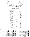
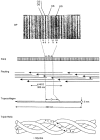
Connective tissues (CTs), which define bodily shape, must respond quickly, robustly and reversibly to deformations caused by internal and external stresses. Fibrillar (elastin, collagen) elasticity under tension depends on molecular and supramolecular mechanisms. A second intra-/inter-molecular pair, involving proteoglycans (PGs), is proposed to cope with compressive stresses. PG interfibrillar bridges ('shape modules'), supramolecular structures ubiquitously distributed throughout CT extracellular matrices (ECMs), are examined for potential elastic properties. L-iduronate residues in shape module decoran PGs are suggested to be molecular springs, cycling through alternative conformations. On a larger scale, anionic glycosaminoglycan (AGAG) interfibrillar bridges in shape modules are postulated to take part in a sliding filament (dashpot-like) process, which converts local compressions into disseminated tensile strains. The elasticity of fibrils and AGAGs, manifest at molecular and larger-scale levels, provides a graduated and smooth response to stresses of varying degrees. NMR and rheo NMR, computer modelling, electron histochemical, biophysical and chemical morphological evidence for the proposals is reviewed.
Figures


Similar articles
-
Cartilage elasticity resides in shape module decoran and aggrecan sumps of damping fluid: implications in osteoarthrosis.J Physiol. 2006 Aug 1;574(Pt 3):643-50. doi: 10.1113/jphysiol.2006.108100. Epub 2006 Mar 31. J Physiol. 2006. PMID: 16581860 Free PMC article.
-
Cartilage is held together by elastic glycan strings. Physiological and pathological implications.Biorheology. 2008;45(3-4):209-17. Biorheology. 2008. PMID: 18836225
-
Structure and function in extracellular matrices depend on interactions between anionic glycosaminoglycans.Pathol Biol (Paris). 2001 May;49(4):284-9. doi: 10.1016/s0369-8114(01)00152-3. Pathol Biol (Paris). 2001. PMID: 11428163 Review.
-
Possible role of decorin glycosaminoglycans in fibril to fibril force transfer in relative mature tendons--a computational study from molecular to microstructural level.J Biomech. 2003 Oct;36(10):1555-69. doi: 10.1016/s0021-9290(03)00133-7. J Biomech. 2003. PMID: 14499303
-
Function of proteoglycans in the extracellular matrix.Acta Pathol Jpn. 1993 Jun;43(6):283-93. doi: 10.1111/j.1440-1827.1993.tb02569.x. Acta Pathol Jpn. 1993. PMID: 8346704 Review.
Cited by
-
Ring-Mesh Model of Proteoglycan Glycosaminoglycan Chains in Tendon based on Three-dimensional Reconstruction by Focused Ion Beam Scanning Electron Microscopy.J Biol Chem. 2016 Nov 4;291(45):23704-23708. doi: 10.1074/jbc.M116.733857. Epub 2016 Sep 13. J Biol Chem. 2016. PMID: 27624935 Free PMC article.
-
Concerted and adaptive alignment of decorin dermatan sulfate filaments in the graded organization of collagen fibrils in the equine superficial digital flexor tendon.J Anat. 2012 Feb;220(2):156-63. doi: 10.1111/j.1469-7580.2011.01456.x. Epub 2011 Nov 28. J Anat. 2012. PMID: 22122012 Free PMC article.
-
Collagen Network Formation in In Vitro Models of Musculocontractural Ehlers-Danlos Syndrome.Genes (Basel). 2023 Jan 24;14(2):308. doi: 10.3390/genes14020308. Genes (Basel). 2023. PMID: 36833235 Free PMC article.
-
The Impact of Post Activation Potentiation on Achilles Tendon Stiffness, Elasticity and Thickness among Basketball Players.Sports (Basel). 2018 Oct 12;6(4):117. doi: 10.3390/sports6040117. Sports (Basel). 2018. PMID: 30321992 Free PMC article.
-
A finite dissipative theory of temporary interfibrillar bridges in the extracellular matrix of ligaments and tendons.J R Soc Interface. 2009 Oct 6;6(39):909-24. doi: 10.1098/rsif.2008.0487. Epub 2008 Dec 23. J R Soc Interface. 2009. PMID: 19106068 Free PMC article.
References
-
- Castellani PP, Moricutti M, Franchi M, Ruggeri A, Roveri N. Arrangement of microfibrils in collagen fibrils in the rat tail. Cell Tiss Res. 1983;234:735–743. - PubMed
-
- Casu B. Discussion. In: Scott JE, editor. Dermatan Sulphate Proteoglycans: Chemistry, Biology, Chemical Pathology. London: The Biochemical Society; 1993. p. p. 76.
-
- Casu B, Petitou M, Provasoli M, Sinay P. Conformational flexibility: a new concept for explaining binding and biological properties of iduronic acid-containing glycosaminoglycans. Trends Biochem Sci. 1988;13:221–225. - PubMed
-
- Davidson EA, Meyer K. Chondroitin, a new mucopolysaccharide. J Biol Chem. 1954;211:605–611. - PubMed
-
- Fatt I. Physiology of the eye. Boston and London: Butterworths; 1978. chap. 6.
Publication types
MeSH terms
Substances
LinkOut - more resources
Full Text Sources
Other Literature Sources
Research Materials

