Review
. 2004 Jan 15;554(Pt 2):285-94.
doi: 10.1113/jphysiol.2003.048439. Epub 2003 Aug 15.

Cell signal control of the G protein-gated potassium channel and its subcellular localization

Affiliations
Review

Cell signal control of the G protein-gated potassium channel and its subcellular localization

Yoshihisa Kurachi et al. J Physiol. .

Abstract

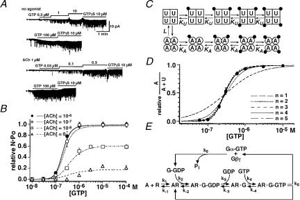
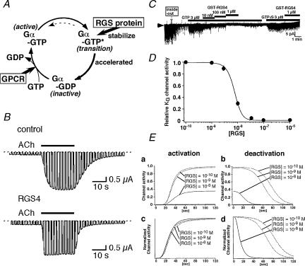
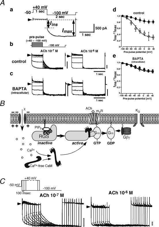
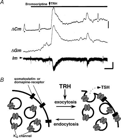
G protein-gated inward rectifier K(+) (K(G)) channels are directly activated by the betagamma subunits released from pertussis toxin-sensitive G proteins, and contribute to neurotransmitter-induced deceleration of heart beat, formation of slow inhibitory postsynaptic potentials in neurones and inhibition of hormone release in endocrine cells. The physiological roles of K(G) channels are critically determined by mechanisms which regulate their activity and their subcellular localization. K(G) channels are tetramers of inward rectifier K(+) (Kir) channel subunits, Kir3.x. The combination of Kir3.x subunits in each K(G) channel varies among tissues and cell types. Each subunit of the channel possesses one Gbetagamma binding site. The binding of Gbetagamma increases the number of functional K(G) channels via a mechanism that can be described by the Monod-Wyman-Changeux allosteric model. During voltage pulses K(G) channel current alters time dependently. The K(G) current exhibits inward rectification due to blockade of outward-going current by intracellular Mg(2+) and polyamines. Upon repolarization, this blockade is relieved practically instantaneously and then the current slowly increases further. This slow current alteration is called 'relaxation'. Relaxation is caused by the voltage-dependent behaviour of regulators of G protein signalling (RGS proteins), which accelerate intrinsic GTP hydrolysis mediated by the Galpha subunit. Thus, the relaxation behaviour of K(G) channels reflects the time course with which the G protein cycle is altered by RGS protein activity at each membrane potential. Subcellular localization of K(G) channels is controlled by several distinct mechanisms, some of which have been recently clarified. The neuronal K(G) channel, which contains Kir3.2c, is localized in the postsynaptic density (PSD) of various neurones including dopaminergic neurones in substantia nigra. Its localization at PSD may be controlled by PDZ domain-containing anchoring proteins. The K(G) channel in thyrotrophs is localized exclusively on secretary vesicles, which upon stimulation are rapidly inserted into the plasma membrane and causes hyperpolarization of the cell. This mechanism indicates a novel negative feedback regulation of exocytosis. In conclusion, K(G) channels are under the control of a variety of signalling molecules which regulate channel activity, subcellular localization and thus their physiological roles in myocytes, neurones and endocrine cells.


Figures

Figure 1
Figure 1. Concentration-dependent effect of intracellular GTP on KG channel
A, examples of inside-out patch experiments obtained from guinea-pig atrial myocytes. The concentration of acetylcholine (ACh) in the pipette was 0 or 1 μm as indicated. The bars above each trace indicate the protocol for application of the various concentrations of GTP and 10 μm GTPγ S to the internal side of the patch membrane. The holding potential was – 80 mV. Note that a 3- to 10-fold increase in GTP concentration resulted in a dramatic increase in channel opening probability (NPo) of KG channels, indicating the existence of a highly cooperative process. B, the relation between the concentration of GTP and the NPo of KG channels normalized to the maximum NPo induced by 10 μm GTPγ S in each patch. Symbols and bars are mean ± s.d. The lines in the graphs are the fits of the data with the Monod–Wyman–Changeux (MWC) allosteric model combined with the Thomsen's model. For fitting the experimental data, only two parameters (k4 and k6) were changed, and best-fitting values were determined by the least-squares method. Other rate constants were kept constant and used the same values as reported by Thomsen et al. (1988). The best-fitting values of each parameter were as follows; k4 = 1.28 × 10⁵ m−1 s−1, k6 = 2.11 × 10⁻² s−1, respectively. C, schematic representation of the MWC allosteric model. In this scheme, each KG channel is assumed to be an oligomer composed of four identical subunits (i.e. n = 4). Each subunit is in either the available (A) or the unavailable (U) state, represented by squares and circles, respectively. Each subunit in the A or U state binds with one dissociated G protein βγ subunit (filled circles) independently of other subunits, with microscopic dissociation constants KA or KU, respectively. In this model, all subunits in the same oligomer must change their conformations simultaneously. Therefore, the channel can be either A4 or U4. A4 and U4 are in equilibrium through an allosteric constant L. D, the fraction of ‘available’ state (A/(A + U)) was calculated from inside-out membrane patch experiments. Lines indicate the fit of the data to the MWC allosteric model with different assumed numbers of n. E, Thomsen's model for receptor–G protein interaction. A, acetylcholine, R, muscarinic m2-receptor, G, G protein. The parameters assayed by biochemical techniques in the Thomsen model are as follows: k1 = 5 × 10⁶ m−1 s−1, k-1 = 0.5 s−1, k2 = 0.1 s−1, k-2 = 0.1 s−1, k3 = 0.1 s−1, k-3 = 1 × 10⁻⁴ s−1, k4 = 1 × 10⁷ m−1 s−1, k-4 = 0.1 s−1, k5 = 0.05 s−1, k6 = 0.10 s−1. Reproduced with permission from Ito et al. (1991) and Hosoya & Kurachi (1999).
Figure 2
Figure 2. RGS protein and KG channel regulation
A, schematic representation of the action of RGS protein. RGS proteins stabilize the transition state (Gα-GTP*) of GTP hydrolysis on the Gα subunit, which results in the acceleration of intrinsic GTPase-activity on the subunit (GTPase-accelerating protein; GAP). GPCR, G protein-coupled receptor. B, whole cell currents through KG channels heterologously composed of Kir3.1/Kir3.4 and m2R were recorded from Xenopus oocytes. Co-injection of RGS4 cRNA (below) accelerated the time courses both of activation and of deactivation. KG currents were evoked by 1 μm ACh. Test pulses of 2 s to –60 mV were applied every 3 s from a holding potential of 0 mV. C, concentration-dependent inhibitory effect of GST-RGS4 on KG currents activated by 3 μm GTPi. The inhibitory effect of 1 μm GST-RGS4 is often hard to wash out, which can be overcome by applying 3 μm GTPγ S. Once irreversibly activated by GTPγ S, KG currents cannot be inhibited by GST-RGS4, suggesting that GTP-hydrolysis reaction is needed for GST-RGS4-mediated inhibition of KG channel activity and that GST-RGS4 may accelerate the intrinsic GTPase-activity of the Gα subunit. D, dose-dependent inhibition of GST-RGS4 on 3 μm GTP-induced KG current. Bars indicate the mean ± s.e.m. The line in the graph is the fit of the data to the MWC allosteric model combined with a modified version of Thomsen's model. For fitting the experimental data, k6 was modified according to eqn (1). The best-fitting values of each parameter were as follows: k6a = 5.6 × 10⁶ m−1 s−1, k6b = 0.024 s−1, respectively. E, the simulated time courses of activation (a and c) and deactivation (b and d) phases of 1 μm ACh-induced KG channel currents in the presence of various concentrations of RGS. RGS not only suppresses maximal channel activity (a and b), it also accelerates the time course of deactivation but not that of activation. Maximal channel activity was normalized in Fig. 2Ec and d. Reproduced with permission from Fujita et al. (2000) and Ishii et al. (2002).
Figure 3
Figure 3. Voltage-dependent ‘relaxation’ property of KG channel
A, effects of extracellular Ca2+ on ACh-induced KG current. Aa, voltage-clamp protocol (upper) and a typical ACh-induced KG current in an isolated atrial myocyte (lower). Inward current upon stepping voltage to –100 mV changes first instantaneously (Iins) and then slowly increases to a steady-state (Imax). Ab, KG current evoked by 10−7 m (left) or 10−6 m (right) of ACh in control conditions. Currents at –100 mV were recorded following prepulses to between –100 and +40 mV in steps of 20 mV Ac, KG current evoked by 10−7 m ACh when intracellular Ca2+ was chelated by BAPTA. Ad and e, relationship between the prepulse voltage and the Iins/Imax ratio for currents elicited by either 10−7 m ACh (open circles) or 10−6 m ACh (filled circles) in control conditions (d) or after intracellular application of BAPTA (e). These results suggest that depolarization-dependent Ca2+ elevation is essential for the agonist concentration-dependent relaxation property of cardiac KG channels. In each current tracen arrowheads indicate the zero current level, and vertical scale bars represent 500 pA. B, schematic representation of voltage-dependent relaxation resulting from Ca2+–CaM-dependent facilitation of the action of RGS proteins. In a hyperpolarized state, the action of RGS is inhibited by PIP3. Once the intracellular Ca2+ concentration is elevated, e.g. upon depolarization, Ca2+–CaM binds to RGS proteins and reverses the inhibitory effect of PIP3, which results in the negative regulation of the G protein cycle. When the Ca2+ concentration decreases to the steady-state level, CaM dissociates from RGS proteins and their action is once again inhibited by PIP3. C, time courses for the depolarization-induced decrease in KG channel availability, which underlies the relaxation resulting from hyperpolarizing voltage steps. Voltage-clamp protocols for envelope pulses (upper) and typical elicited currents in the presence of 0.1 μm ACh (lower left) or 1 μm ACh (lower right) are shown. Reproduced with permission from Ishii et al. (2001).
Figure 4
Figure 4. Secretagogue-induced cell surface recruitment of KG channel in thyrotroph
A, simultaneous recordings of Cm (membrane capacitance), Gm (membrane conductance) and Im (whole cell current) during stimulation of a thyrotroph cell. Although the application of bromocriptine to the bath did not induce any increase in Cm, the addition of TRH clearly increased Cm. Cm returned gradually to near basal level after TRH application (upper trace). Im was slightly changed by bromocriptine alone. Addition of TRH induced a marked increase in the inward Im at the holding potential of –100 mV. The arrowhead indicates the zero current level. Filled circles show calibration signals for Cm (250 fF). The scale bar for Cm is 250 fF, which corresponds to 2.35 nS, and the scale bar for Im is 250 pA. Time scale bar = 1 min. B, a schematic illustration of TRH-induced recruitment of KG channels via exocytotic fusion. The agonist-induced K+ channel activation would thus be augmented by the TRH-induced exocytosis. Reproduced with permission from Morishige et al. (1999).

Similar articles

Cited by

References

    1. Breitwieser GE, Szabo G. Uncoupling of cardiac muscarinic and β-adrenergic receptors from ion channels by a guanine nucleotide analogue. Nature. 1985;317:538–540. - PubMed
    1. Burgen ASV, Terroux KG. On the negative inotropic effect in the cat's auricle. J Physiol. 1953;120:449–464. - PMC - PubMed
    1. Cohen NA, Brenman JE, Snyder SH, Bredt DS. Binding of the inward rectifier K+ channel Kir 2.3 to PSD-95 is regulated by protein kinase A phosphorylation. Neuron. 1996;17:759–767. - PubMed
    1. Corey S, Clapham DE. The Stoichiometry of Gβγ binding to G-protein-regulated inwardly rectifying K+ channels (GIRKs) J Biol Chem. 2001;276:11409–11413. - PubMed
    1. Dascal N, Lim NF, Schreibmayer W, Wang W, Davidson N, Lester HA. Expression of an atrial G-protein-activated potassium channel in Xenopus oocytes. Proc Natl Acad Sci USA. 1993;90:6569–6600. - PMC - PubMed

Publication types

MeSH terms

Substances

LinkOut - more resources