. 2003 Sep;9(9):1123-37.
doi: 10.1261/rna.5610403.

Partitioning and translation of mRNAs encoding soluble proteins on membrane-bound ribosomes

Affiliations

Partitioning and translation of mRNAs encoding soluble proteins on membrane-bound ribosomes

Rachel S Lerner et al. RNA. 2003 Sep.

Abstract

In eukaryotic cells, it is generally accepted that protein synthesis is compartmentalized; soluble proteins are synthesized on free ribosomes, whereas secretory and membrane proteins are synthesized on endoplasmic reticulum (ER)-bound ribosomes. The partitioning of mRNAs that accompanies such compartmentalization arises early in protein synthesis, when ribosomes engaged in the translation of mRNAs encoding signal-sequence-bearing proteins are targeted to the ER. In this report, we use multiple cell fractionation protocols, in combination with cDNA microarray, nuclease protection, and Northern blot analyses, to assess the distribution of mRNAs between free and ER-bound ribosomes. We find a broad representation of mRNAs encoding soluble proteins in the ER fraction, with a subset of such mRNAs displaying substantial ER partitioning. In addition, we present evidence that membrane-bound ribosomes engage in the translation of mRNAs encoding soluble proteins. Single-cell in situ hybridization analysis of the subcellular distribution of mRNAs encoding ER-localized and soluble proteins identify two overall patterns of mRNA distribution in the cell-endoplasmic reticular and cytosolic. However, both partitioning patterns include a distinct perinuclear component. These results identify previously unappreciated roles for membrane-bound ribosomes in the subcellular compartmentalization of protein synthesis and indicate possible functions for the perinuclear membrane domain in mRNA sorting in the cell.


Figures
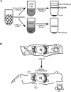
FIGURE 1.
FIGURE 1.
Methods for fractionation of cultured cells. (A) Mechanical homogenization. Tissue culture cells were suspended in hypotonic buffer, homogenized, and either mixed with high density (2.5 M) sucrose and centrifuged in a discontinuous gradient to yield isopycnic separation of the endoplasmic reticulum fraction, or subjected to differential velocity sedimentation to yield a cytosol fraction. (B) Detergent permeabilization. Tissue culture cells were treated with digitonin, at concentrations sufficient to yield permeabilization of the plasma membrane, without disruption of the integrity of the intracellular organelles. The released cytosol fraction, containing the free polysome fraction, was separated from the endoplasmic-reticulum-bound fraction by differential centrifugation.
FIGURE 2.
FIGURE 2.
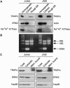
Marker protein analysis of subcellular fractions obtained by mechanical homogenization or detergent-based fractionation of Jurkat or J558 cells. (A) Samples of homogenate, cytosol, or purified ER membrane fractions, obtained as depicted in Figure 1A ▶, were separated by SDS-PAGE, transferred to nitrocellulose membranes, and analyzed for the ER membrane marker TRAPα, the cytosol markers ERK2 or actin, or the plasma membrane marker Na+/K+ ATPase, by immunoblot analyses. (B) Subcellular fractions, as in A, were screened for rRNA and tRNA by denaturing RNA gel analysis. (C) Total cell extracts, cytosol (detergent-releasable), or membrane-bound (detergent-resistant) fractions obtained as in Figure 1B ▶ were separated on SDS-PAGE and analyzed for marker protein content (TRAPα, ERK2, or Hsp90) by immunoblot.
FIGURE 3.
FIGURE 3.
Electron micrographic analysis of digitonin-based cell fractionation. J558 murine plasmacytoma cells were fixed in glutaraldehyde and processed for thin-section electron microscopy either (A) prior to or (B) following digitonin extraction of the cytosol contents. Low-magnification images of control (A) or digitonin-extracted cells (B) are depicted. Bar, 3 μm. (CF) High magnification representative sections of (C) the cytosol/ER membrane junction and (D) an isolated ER membrane profile of control cells; (E,F) similar, representative sections of digitonin-extracted cells. For both sections, (arrowheads) ribosome profiles, (arrows) ER luminal aggregates, and (*) ER luminal contents. Bar, 0.5 μm.
FIGURE 4.
FIGURE 4.
Steady-state mRNA partitioning between cytosolic and ER-bound polysomes. Total RNA was isolated from cytosol and ER membrane fractions of Jurkat and J558 cells, as indicated in Figure 1A ▶. For each reaction, 10 μg of RNA was incubated with one of eight [32P]-labeled oligonucleotide probes representing three different classes of genes, based on the type of protein encoded: cytosolic, ER membrane, and ER lumenal. The nuclease-protected RNA fragments from parallel reactions with cytosol and ER-derived RNA were purified and separated by denaturing acrylamide gel electrophoresis. Quantification was performed by phosphorimager analysis; photostimulated luminescence output (PSL) values are indicated. Digital images of the phosphorimager output are depicted.
FIGURE 5.
FIGURE 5.
Analysis by cDNA microarray of mRNA distribution between free and ER-membrane-bound polysomes of Jurkat cells. Total and ER-membrane-bound RNA fractions were obtained from Jurkat cells, as described in the legend to Figure 1A ▶, and used to generate probes for cDNA microarray analysis. Stratagene 1.2K microarrays were used, with scoring performed as described in Materials and Methods. (A) The absolute display of total mRNAs in the ER-bound polysome fraction. (B) The relative enrichment of those mRNAs significantly represented in the total RNA fraction, and present in the ER-bound polysome fraction. Depicted also are the number of mRNAs displaying marked (>fourfold) enrichment or depletion in the ER polysome fraction.
FIGURE 6.
FIGURE 6.
Single-cell in situ hybridization analysis of mRNA distribution in intact cells. NIH-3T3 cells were fixed and processed for in situ hybridization, as described in Materials and Methods. Cells were probed with digoxygenin-labeled riboprobes against mRNAs encoding (A) the ER-resident protein GRP94, and the cytosolic proteins (B) GAPDH and (C) Hsp90. Detection was performed using alkaline phosphatase conjugated anti-digoxygenin antibodies, and Vector Stain Red alkaline phosphatase substrate. Three representative images from each analysis are depicted. Digital images were captured and probe distribution was rendered in 3D surface projections using ImageJ software.
FIGURE 7.
FIGURE 7.
Perinuclear localization of GAPDH mRNAs in NIH-3T3 cells. NIH-3T3 cells were processed for single-cell in situ hybridization analyses of mRNA encoding GAPDH, as described in the legend to Figure 6 ▶. Optical sections (A,B) nearing the central nuclear plane and (C,D) through the central nuclear plane are depicted. The distinct perinuclear staining evident for a subpopulation of the GAPDH mRNA is highlighted in these discrete sections.
FIGURE 8.
FIGURE 8.
Membrane-bound ribosomes are engaged with mRNAs encoding cytosolic proteins. (A) Analysis of mRNA distribution in the cell fractions obtained by detergent fractionation. Total cell (T), membrane-bound (B), and cytosolic, or free (F), fractions were obtained by digitonin extraction of Jurkat cells, as depicted in the legend to Figure 1B ▶. Samples of 10 μg of total RNA were separated on agarose denaturing gels and transferred to nitrocellulose membranes. Following staining to confirm RNA integrity, blots were processed for Northern analysis using probes directed against mRNAs encoding the ER-resident proteins GRP94 and BiP and the cytosol proteins GAPDH and Hsp90. (B) mRNAs encoding cytosolic proteins are represented on ER-membrane-bound polysomes. Jurkat cells were treated with cycloheximide to stabilize polysome structure and fractionated, as depicted in Figure 1B ▶, to yield the ER-membrane-bound polysome fraction. The ER polysome fraction was solubilized in detergent and fractionated by velocity sedimentation on 15%–50% sucrose gradients. Gradients were fractionated and analyzed by UV spectrometry. RNA was extracted from each fraction for Northern blot analysis, against GAPDH and Hsp90, and to identify rRNAs (18S, 28S) representing the small and large ribosomal subunits. (C,D) mRNAs maintain association with ribosomes. Jurkat cells were treated with (C) cycloheximide to maintain polysome structure or with (D) pactamycin to cause run-off translation and elicit polysome breakdown. Cells were then harvested and lysed, and polysome profiles were attained by the methods described in B. The RNA contained in each gradient fraction was analyzed by nuclease protection assay, as described in Figure 4 ▶.

References

    1. Adam, S.A., Marr, R.S., and Gerace, L. 1990. Nuclear protein import in permeabilized mammalian cells requires soluble cytoplasmic factors. J. Cell Biol. 111: 807–816. - PMC - PubMed
    1. Bassell, G.J., Oleynikov, Y., and Singer, R.H. 1999. The travels of mRNAs through all cells large and small. FASEB J. 13: 447–454. - PubMed
    1. Blobel, G. and Dobberstein, B. 1975a. Transfer of proteins across membranes. I. Presence of proteolytically processed and unprocessed nascent immunoglobulin light chains on membrane-bound ribosomes of murine myeloma. J. Cell Biol. 67: 835–851. - PMC - PubMed
    1. ———. 1975b. Transfer of proteins across membranes. II. Reconstitution of functional rough microsomes from heterologous components. J. Cell Biol. 67: 852–862. - PMC - PubMed
    1. Brooks, J.C. and Treml, S. 1984. Effect of trifluoperazine and calmodulin on catecholamine secretion by saponin-skinned cultured chromaffin cells. Life Sci. 34: 669–674. - PubMed

Publication types

LinkOut - more resources