. 2003 Sep;9(9):1157-67.
doi: 10.1261/rna.5720303.

An endonuclease activity similar to Xenopus PMR1 catalyzes the degradation of normal and nonsense-containing human beta-globin mRNA in erythroid cells

Affiliations

An endonuclease activity similar to Xenopus PMR1 catalyzes the degradation of normal and nonsense-containing human beta-globin mRNA in erythroid cells

Kirsten A Bremer et al. RNA. 2003 Sep.

Abstract

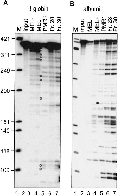
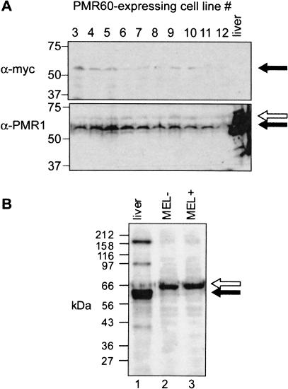
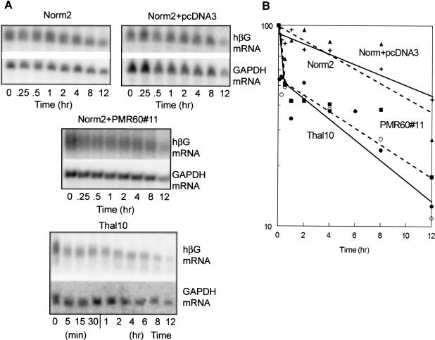
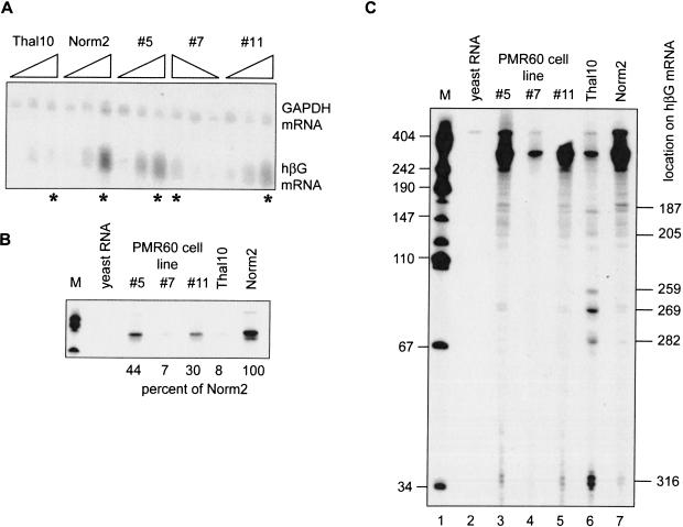
beta-globin mRNA bearing a nonsense codon is degraded in the cytoplasm of erythroid cells by endonuclease cleavage, preferentially at UG dinucleotides. An endonuclease activity in polysomes of MEL cells cleaved beta-globin and albumin mRNA in vitro at many of the same sites as PMR1, an mRNA endonuclease purified from Xenopus liver. Stable transfection of MEL cells expressing normal human beta-globin mRNA with a plasmid vector expressing the catalytically active form of PMR1 reduced the half-life of beta-globin mRNA from 12 to 1-2 h without altering GAPDH mRNA decay. The reduced stability of beta-globin mRNA in these cells was accompanied by an increase in the production of mRNA decay products corresponding to those seen in the degradation of nonsense-containing beta-globin mRNA. Therefore, beta-globin mRNA is cleaved in vivo by an endonuclease with properties similar to PMR1. Inhibiting translation with cycloheximide stabilized nonsense-containing beta-globin mRNA, resulting in a fivefold increase in its steady-state level. Taken together, our results indicate that the surveillance of nonsense-containing beta-globin mRNA in erythroid cells is a cytoplasmic process that functions on translating mRNA, and endonucleolytic cleavage constitutes one step in the process of beta-globin mRNA decay.


Figures

FIGURE 1.
FIGURE 1.
In vitro cleavage of β-globin and albumin mRNA by PMR1 and the β-globin mRNA endonuclease. (A) A 5′ 32P-labeled transcript of the 5′-most 347 nucleotides of human β-globin mRNA was incubated for 30 min at 37°C with no added protein (lane 2, input), polysome extract from uninduced (MEL−, lane 3) or 48-h DMSO-induced MEL cells (MEL+, lane 4), or two fractions containing β-globin mRNA endonuclease activity recovered from Mono S fractionation of the polysome extract (lanes 6,7). Alternatively, the transcript was incubated for 20 min at 25°C with 20 U of purified Xenopus PMR1 (PMR1, lane 5). The reaction products were separated on a denaturing 6% polyacrylamide/urea gel and visualized by PhosphorImager. Lane 1 contains a marker consisting of φX174 DNA HinfI fragments and the open circles identify products with counterparts in vivo (Stevens et al. 2002). (B) An in vitro-synthesized, 5′ 32P-labeled transcript consisting of the 5′-most 420 nucleotide of Xenopus albumin mRNA was incubated as described in A. The characteristic PMR1 cleavage at overlapping APyrUGA elements is identified by a solid circle.
FIGURE 2.
FIGURE 2.
Expression of myc-PMR60 in transfected Norm2 cells. (A) Norm2 cells were stably transfected with a plasmid expressing the catalytically active 60-kD form of Xenopus PMR1 bearing an amino-terminal myc epitope tag. Equal amounts of cytoplasmic extract from 10 stable cell lines recovered following selection were applied to duplicate 10% SDS-PAGE gels. In addition, one blot contained a sample of Xenopus liver polysome extract. Western blots of the duplicate gels were probed with a monoclonal antibody to the myc epitope (top) or a polyclonal antibody to Xenopus PMR1 (Dompenciel et al. 1995) (bottom). The solid arrow corresponds to a 60-kD PMR1, and the open arrow to a larger cross-reacting protein seen in all cultures. (B) Polysomes from Xenopus liver (lane 2), uninduced MEL cells (lane 2, MEL−), or DMSO-induced MEL cells (lane 3, MEL+) were analyzed as above by Western blotting using a polyclonal antibody to Xenopus PMR1.
FIGURE 3.
FIGURE 3.
β-globin mRNA turnover in Norm2 cells, Thal10 cells, or Norm2 cells transfected with vector or PMR1. (A) Norm2 cells expressing wild-type β-globin mRNA (Norm2), Thal10 cells expressing nonsense-containing β-globin mRNA (Thal10), Norm2 cells that were stably transfected with empty vector (Norm2+pcDNA3), or a line of Norm2 cells stably transfected with vector expressing PMR1 (Norm2+PMR60#11) were induced for 48 h with DMSO prior to addition of actinomycin D. Cytoplasmic RNA was isolated at the indicated times and analyzed by Northern blot for human β-globin mRNA (hβG, top, each set) or GAPDH mRNA (bottom, each set). Shown are typical results from three independent experiments. The results in A were quantified by PhosphorImager analysis and are presented graphically in B. With the exception of Thal10 cells (•, ▪, solid line) the datapoints represent the decay of the mRNAs in A (Norm2 +, solid line; Norm2+pcDNA3 ▴, broken line; Norm2+PMR60#11 ○, broken line. The data points for Thal10 cells consist of those derived from the blot in A plus an independent experiment (not shown).
FIGURE 4.
FIGURE 4.
Identification of β-globin mRNA decay intermediates in Norm2 cells expressing Xenopus PMR1. Cytoplasmic RNA was isolated at time 0, 48, and 96 h following DMSO-induction from Norm2, Thal10, or three lines of Norm2 cells stably transfected with plasmids expressing myc-PMR60. (A) Each of the RNA samples was analyzed by Northern blot that was hybridized to a mixed probe for β-globin and GAPDH mRNA. The RNAs were applied in the order indicated above with the exception of cell line #7, in which they were applied in reverse order. (B) Equal amounts of the 96-h samples identified with by an asterisk (*) in A were analyzed by S1 nuclease protection using a NaeI–BamH1 probe, and the protected products were analyzed as described previously (Stevens et al. 2002). An 18-h exposure of the portion of the gel corresponding to the fully protected probe is shown in B. The marker (M) in lane 1 consists of HpaII fragments of pUC19 DNA. (Lane 2) Yeast RNA was added to the initial hybridization reaction; (lanes 3–5) S1 nuclease protection was performed on RNA obtained from three independent clones of Norm2 cells stably transfected with myc-PMR60; (lanes 6,7) S1 nuclease protection was performed on RNAs from Thal10 and parental Norm2 cells. The relative quantity of each fully protected product was determined by scanning densitometry, and the percent of the signal for β-globin mRNA from Norm2 cells is shown beneath the gel. (C) The gel was exposed for 96 h to visualize β-globin mRNA decay intermediates. Human β-globin mRNA decay products characterized previously (Stevens et al. 2002) are identified on the right side of the autoradiogram.
FIGURE 5.
FIGURE 5.
Impact of inhibiting translation on steady-state levels of human β-globin mRNA in transfected MEL cells. The MEL cell transfectants described in the legend to Figure 4 ▶ were induced with DMSO for 48 h. At time 0, the medium the cells either received fresh medium (A) or medium containing cycloheximide (B). Cytoplasmic RNA was isolated at the indicated time points and analyzed by Northern blot for human β-globin mRNA (top, each set) and GAPDH (bottom, each set).
FIGURE 6.
FIGURE 6.
Impact of inhibition translation mRNA on turnover in Norm2 cells, Thal10 cells, or Norm2 cells transfected with vector or PMR1. The MEL cell transfectants described in Figure 4 ▶ were induced with DMSO for 48 h. Cycloheximide was added at time 0 (0) and RNA was isolated 4 h later to monitor the impact on inhibiting translation on mRNA steady state levels (+CHX). Actinomycin D was added at this point and cytoplasmic RNA was isolated at the indicated times over the next 8 h. The isolated RNAs were analyzed by Northern blot for human β-globin mRNA (top, each set) and GAPDH mRNA (bottom, each set).

Similar articles

Cited by

References

    1. Antoniou, M. 1991. Induction of erythroid-specific expression in murine erythroleukemia (MEL) cell line. In Methods in molecular biology. Gene transfer and expression protocols (ed. E.J. Murray), pp. 421–434. Humana Press, Clifton, NJ. - PubMed
    1. Antoniou, M. and Grosveld, F. 1990. β-globin dominant control region interacts differently with distal and proximal promoter elements. Genes & Dev. 4: 1007–1013. - PubMed
    1. Bandyopadhyay, R., Coutts, M., Krowczynska, A., and Brawerman, G. 1990. Nuclease activity associated with mammalian mRNA in its native state: Possible basis for selectivity in mRNA decay. Mol. Cell. Biol. 10: 2060–2069. - PMC - PubMed
    1. Bashkirov, V.I., Scherthan, H., Solinger, J.A., Buerstedde, J.M., and Heyer, W.D. 1997. A mouse cytoplasmic exoribonuclease (mXRN1p) with preference for G4 tetraplex substrates. J. Cell Biol. 136: 761–773. - PMC - PubMed
    1. Binder, R., Horowitz, J.A., Basilion, J.P., Koeller, D.M., Klausner, R.D., and Harford, J.B. 1994. Evidence that the pathway of transferrin receptor mRNA degradation involves an endonucleolytic cleavage within the 3′ UTR and does not involve poly(A) tail shortening. EMBO J. 13: 1969–1980. - PMC - PubMed

Publication types

LinkOut - more resources