. 2003 Sep 30;100 Suppl 1(Suppl 1):11836-41.
doi: 10.1073/pnas.1734197100. Epub 2003 Aug 15.

Screening for mammalian neural genes via fluorescence-activated cell sorter purification of neural precursors from Sox1-gfp knock-in mice

Affiliations

Screening for mammalian neural genes via fluorescence-activated cell sorter purification of neural precursors from Sox1-gfp knock-in mice

Jerome Aubert et al. Proc Natl Acad Sci U S A. .

Abstract

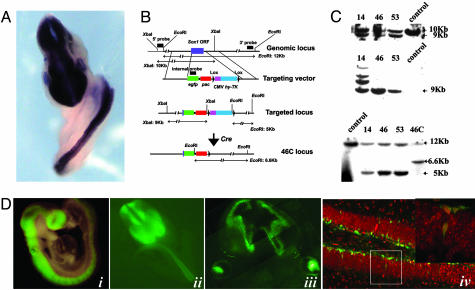
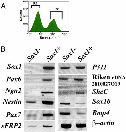
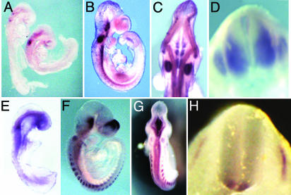
The transcription factor Sox1 is the earliest and most specific known marker for mammalian neural progenitors. During fetal development, Sox1 is expressed by proliferating progenitor cells throughout the central nervous system and in no tissue but the lens. We generated a reporter mouse line in which egfp is inserted into the Sox1 locus. Sox1GFP animals faithfully recapitulate the expression of the endogenous gene. We have used the GFP reporter to purify neuroepithelial cells by fluorescence-activated cell sorting from embryonic day 10.5 embryos. RNAs prepared from Sox1GFP+ and Sox1GFP- embryo cells were then used to perform a pilot screen of subtracted cDNAs prepared from differentiating embryonic stem cells and arrayed on a glass chip. Fifteen unique differentially expressed genes were identified, all previously associated with fetal or adult neural tissue. Whole mount in situ hybridization against two genes of previously unknown embryonic expression, Lrrn1 and Musashi2, confirmed the selectivity of this screen for early neuroectodermal markers.


Figures

Fig. 1.
Fig. 1.
Targeting Sox1 and Sox1GFP expression. (A) In situ hybridization of an E10.5 embryo with a Sox1 riboprobe, showing expression restricted to and throughout the neuraxis. (B) Schematic showing the design of the targeting vector and the screening strategy for identification of correctly targeted clones. (C) Southern blots showing the correct targeting events and the excision of the cytomegalovirus (CMV) HyTK cassette after transient transfection with a Cre expression plasmid. (Top)5′ probe. (Middle) Internal EGFP probe. (Bottom)3′ probe. Clone 14 is correctly targeted but has multiple integrations; clones 46 and 53 are correctly targeted, single integration clones. Clone 46C is a derivative of clone 46 after Cre-mediated deletion of the CMV Hy-TK cassette. Control is DNA from the parental E14Tg2a ES cells. (D) Sox1GFP expression in embryos and adult animals. (i) Sox1GFP embryo at E9.5 showing expression throughout the length of the neural tube. (ii) Dorsal anterior view of a Sox1GFP embryo at E11.5 showing exclusion of GFP fluorescence from the midline, indicating no expression in roofplate and floorplate. (iii) Coronal section through the head of an E12.5 embryo showing Sox1GFP expression in the ventricular and subventricular zone and the lens. (iv) Section through an adult Sox1GFP mouse brain, showing Sox1GFP-expressing cells in the subgranular layer of the hippocampus. (Inset) Higher magnification of boxed area, showing a GFP-expressing cell in the granular layer.
Fig. 2.
Fig. 2.
FACS purification of neural precursors. (A) FACS profile showing cell sorting of the Sox1+ neural precursor population from the Sox1-cell population from E10.5 mouse embryos. (B) Positive (R2, 30.35%) and negative (R1, 44.02%) populations were sorted and analyzed by RT-PCR for marker gene expression.
Fig. 3.
Fig. 3.
Expression of Lrrn1 and Musashi2 mRNAs in mouse embryos. (A) Lateral view of E8.5 embryo showing Msi2h hybridization in the hindbrain and the otic vesicle. (B and C) Lateral and dorsal view, respectively, of an E10.5 embryo showing Msi2h hybridization in the neural tube, hindbrain, and otic vesicles. (D) Transverse section at E11.5 showing specific Msi2h hybridization in the ventral half of the neural tube and the dorsal root ganglia. (E) Lateral view of an E8.5 embryo showing Lrrn1 hybridization in the neural tube and weakly in the somites. (F and G) Lateral and dorsal view, respectively, of an E10.5 embryo showing Lrrn1 hybridization in the telencephalic vesicle, the hindbrain, the otic vesicle, and the somites. (H) Transverse section at E10.5 shows Lrrn1 hybridization in the ventral part of the neural tube and the somites.

References

    1. Rossi, F. & Cattaneo, E. (2002) Nat. Rev. Neurosci. 3, 401-409. - PubMed
    1. Gorba, T. & Allsopp, T. (2003) Pharmacol. Res. 47, 269-278. - PubMed
    1. Svendsen, C. N. & Smith, A. G. (1999) Trends Neurosci. 22, 357-364. - PubMed
    1. Evans, M. J. & Kaufman, M. H. (1981) Nature 292, 154-156. - PubMed
    1. Doetschman, T. C., Eistetter, H., Katz, M., Schmidt, W. & Kemler, R. (1985) J. Embryol. Exp. Morphol. 87, 27-45. - PubMed

Publication types

MeSH terms