

Detecting microdamage in bone
- PMID: 12924817
- PMCID: PMC1571153
- DOI: 10.1046/j.1469-7580.2003.00211.x
Detecting microdamage in bone
Abstract
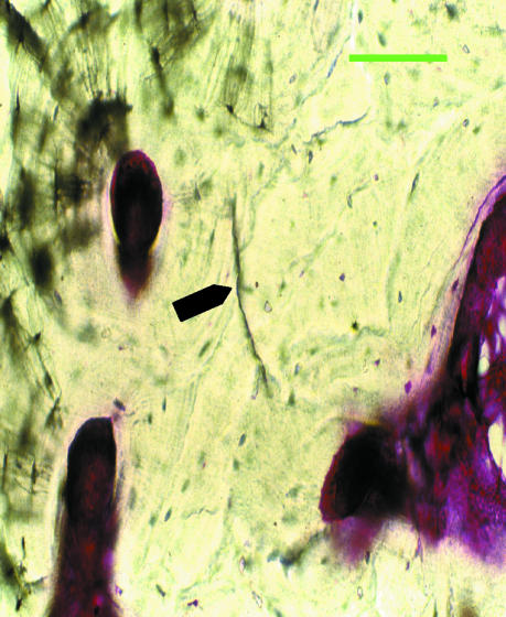
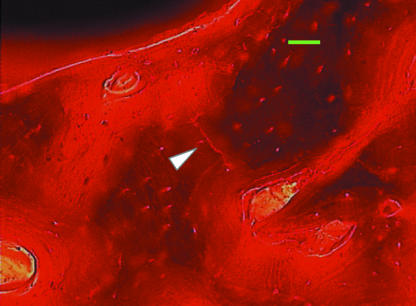
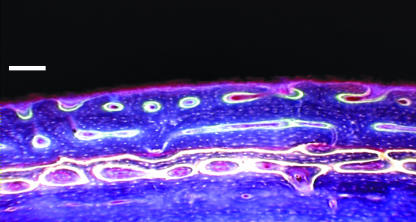
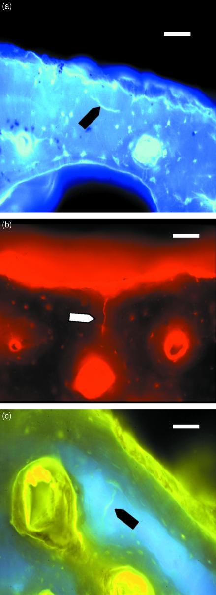
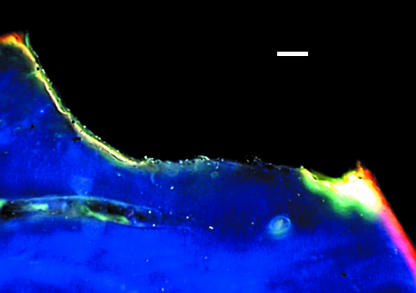
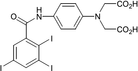
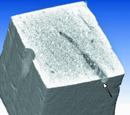
Fatigue-induced microdamage in bone contributes to stress and fragility fractures and acts as a stimulus for bone remodelling. Detecting such microdamage is difficult as pre-existing microdamage sustained in vivo must be differentiated from artefactual damage incurred during specimen preparation. This was addressed by bulk staining specimens in alcohol-soluble basic fuchsin dye, but cutting and grinding them in an aqueous medium. Nonetheless, some artefactual cracks are partially stained and careful observation under transmitted light, or epifluorescence microscopy, is required. Fuchsin lodges in cracks, but is not site-specific. Cracks are discontinuities in the calcium-rich bone matrix and chelating agents, which bind calcium, can selectively label them. Oxytetracycline, alizarin complexone, calcein, calcein blue and xylenol orange all selectively bind microcracks and, as they fluoresce at different wavelengths and colours, can be used in sequence to label microcrack growth. New agents that only fluoresce when involved in a chelate are currently being developed--fluorescent photoinduced electron transfer (PET) sensors. Such agents enable microdamage to be quantified and crack growth to be measured and are useful histological tools in providing data for modelling the material behaviour of bone. However, a non-invasive method is needed to measure microdamage in patients. Micro-CT is being studied and initial work with iodine dyes linked to a chelating group has shown some promise. In the long term, it is hoped that repeated measurements can be made at critical sites and microdamage accumulation monitored. Quantification of microdamage, together with bone mass measurements, will help in predicting and preventing bone fracture failure in patients with osteoporosis.
Figures
Similar articles
-
Fluorescence-aided detection of microdamage in compact bone.J Anat. 1998 Aug;193 ( Pt 2)(Pt 2):179-84. doi: 10.1046/j.1469-7580.1998.19320179.x. J Anat. 1998. PMID: 9827633 Free PMC article.
-
Sequential labelling of microdamage in bone using chelating agents.J Orthop Res. 2000 Mar;18(2):322-5. doi: 10.1002/jor.1100180222. J Orthop Res. 2000. PMID: 10815835 Clinical Trial.
-
New insights into the propagation of fatigue damage in cortical bone using confocal microscopy and chelating fluorochromes.Eur J Morphol. 2005 Feb-Apr;42(1-2):81-90. doi: 10.1080/09243860500096206. Eur J Morphol. 2005. PMID: 16123027
-
[Morphological analysis of bone dynamics and metabolic bone disease. Pathophisiology of Microdamage].Clin Calcium. 2011 Apr;21(4):559-65. Clin Calcium. 2011. PMID: 21447923 Review. Japanese.
-
Microdamage and bone mechanobiology.Technol Health Care. 2006;14(4-5):359-65. Technol Health Care. 2006. PMID: 17065757 Review.
Cited by
-
Trabecular bone microcrack accumulation in patients treated with bisphosphonates for durations up to 16 years.J Orthop Res. 2023 May;41(5):1033-1039. doi: 10.1002/jor.25441. Epub 2022 Sep 26. J Orthop Res. 2023. PMID: 36163612 Free PMC article.
-
Synchrotron radiation micro-CT at the micrometer scale for the analysis of the three-dimensional morphology of microcracks in human trabecular bone.PLoS One. 2011;6(7):e21297. doi: 10.1371/journal.pone.0021297. Epub 2011 Jul 7. PLoS One. 2011. PMID: 21750707 Free PMC article.
-
Modalities for Visualization of Cortical Bone Remodeling: The Past, Present, and Future.Front Endocrinol (Lausanne). 2015 Aug 11;6:122. doi: 10.3389/fendo.2015.00122. eCollection 2015. Front Endocrinol (Lausanne). 2015. PMID: 26322017 Free PMC article. Review.
-
Translational aspects of bone quality--vertebral fractures, cortical shell, microdamage and glycation: a tribute to Pierre D. Delmas.Osteoporos Int. 2009 May;20 Suppl 3:S247-53. doi: 10.1007/s00198-008-0791-z. Osteoporos Int. 2009. PMID: 19430876 Review.
-
Microdamage as a Bone Quality Component: Practical Guidelines for the Two-Dimensional Analysis of Linear Microcracks in Human Cortical Bone.JBMR Plus. 2019 Jul 17;3(6):e10203. doi: 10.1002/jbm4.10203. eCollection 2019 Jun. JBMR Plus. 2019. PMID: 31346569 Free PMC article. Review.
References
-
- Aaron JE, Makins NB, Francis RB, Peacock M. Staining of the calcification front in human bone using contrasting fluorochromes in vitro. J. Histochem. Cytochem. 1984;32:1251–1261. - PubMed
-
- Akhter MP, Taber BJ, Kimmel DB, Recker RR. Demonstration of fatigue damage in rat tibiae. Proc. Orthop. Res. Soc. 1993;39:197.
-
- Andre T. Studies on the distribution of tritium-labelled dihydrostreptomycin and tetracycline in the body. Acta Radiolog. Suppl. 1956;142:1–90. - PubMed
-
- Bentolila V, Boyce TM, Fyhrie DP, Drumb R, Skerry TM, Schaffler MB. Intracortical remodelling in adult rat long bones after fatigue loading. Bone. 1998;23:275–281. - PubMed
-
- Blackwood RK. Structure determination and total synthesis of the tetracyclines. In: Hlavka JJ, Boothe JH, editors. The Tetracyclines. Handbook of Experimental Pharmacology. Vol. 78. Berlin: Springer-Verlag; 1985. pp. 85–90.
Publication types
MeSH terms
Substances
Grants and funding
LinkOut - more resources
Full Text Sources
Other Literature Sources
Medical
