Review
. 2003 Aug;203(2):173-89.
doi: 10.1046/j.1469-7580.2003.00213.x.

The real response of bone to exercise

Affiliations
Review

The real response of bone to exercise

Alan Boyde. J Anat. 2003 Aug.

Abstract

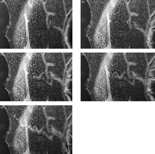
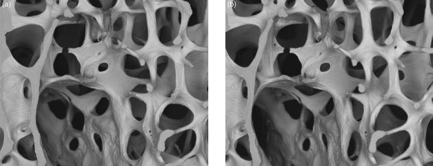
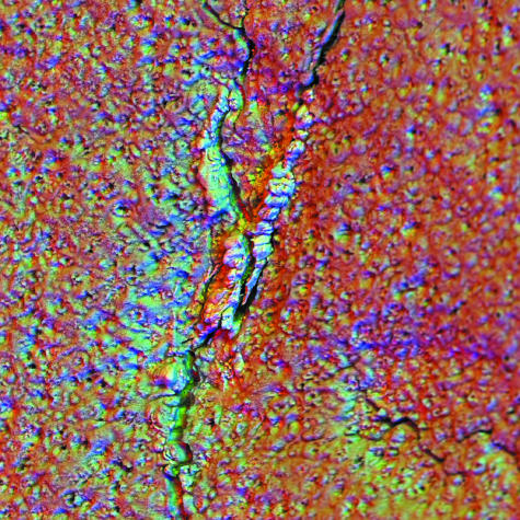
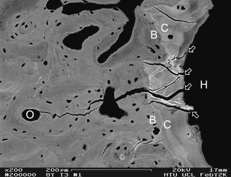
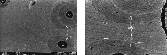
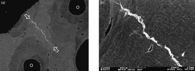
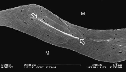
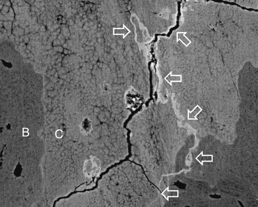
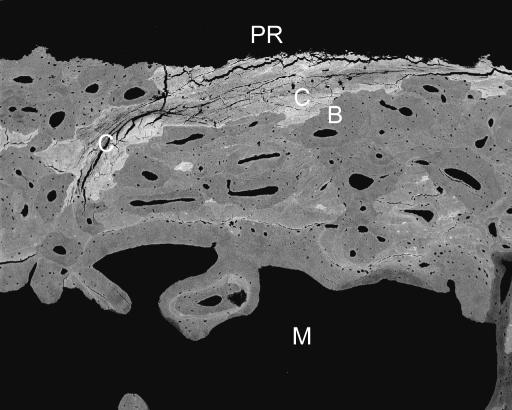
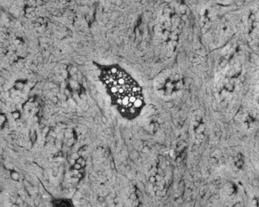
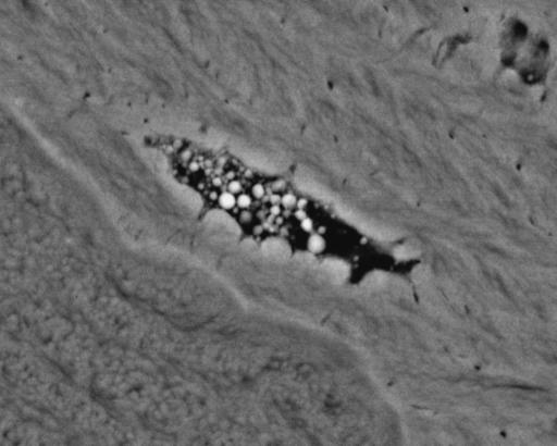
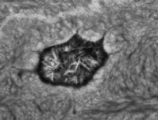
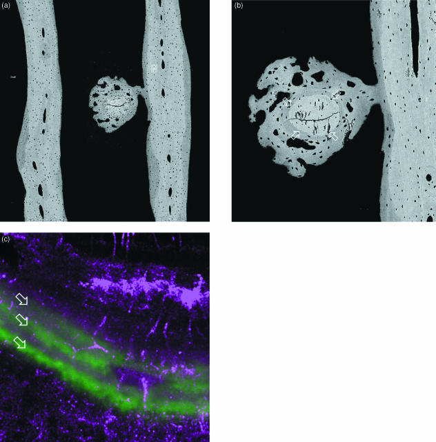
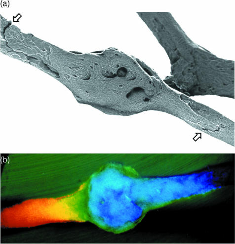
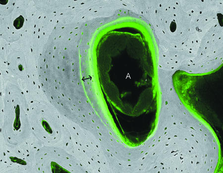
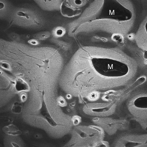
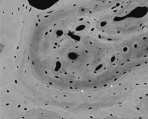
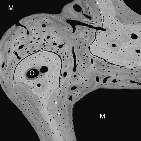
This review presents findings made in studies of large mammalian bones, especially from racehorse training experiments (2-8 years old, third metacarpal, tarsal) and human autopsy orthopaedic femoral implant retrievals and other human biopsy and autopsy cases. Samples were cleaned to analyse mineralized matrix in three dimensions, or poly methyl-methacrylate embedded and micromilled to delete topography and study the superficial c. 0.5-microm two-dimensional section using quantitative backscattered electron imaging. With experimental implant studies in rabbits, observations were also made in vivo using confocal microscopy. Cracks in both calcified cartilage and bone may be removed by infilling with calcified matrix. This may be a general repair mechanism for calcified connective tissue crack repair. The fraction of the organ volume occupied by any form of bone tissue in equine distal third metacarpal extremities was increased in the more exercised groups by bone deposited within former marrow adipocytic space. Where deposited upon prior lamellar bone surfaces, this occurred without the intervention of prior resorption and without the formation of a hypermineralized cement line. Exercise inhibited osteoclastic resorption at external anatomical growth modelling sites where it normally occurs. Addition is not coupled to time-wasting resorption: both internally and externally, it occurs both by layering on existing cancellous surfaces and by creation of new immature scaffold, with de novo incorporation of a rich, capillary blood vessel supply. The real response within bone organs subjected to mechanical overload exercise within normal physiological limits is to make more, and to lose less, bone.


Figures

Fig. 1
Fig. 1
Dynamic 3D LM study of fracture initiation and propagation in isolated equine Mc3 trabeculae. One side of stereo-pair images. 10× water-immersion objective, Edge 3D microscope.
Fig. 2
Fig. 2
20-kV stereo pair BSE image of three-line bending test sample of fourth lumbar vertebral body trabecular bone, showing the very complex fracture path. Tilt angle difference 8°. Field width, 4.45 mm.
Fig. 3
Fig. 3
Medial condylar groove of 2-year-old Thoroughbred racehorse from MUGES training study, hyaline articular cartilage removed to the level of the mineralizing front (MF) of ACC using Tergazyme, carbon-coated, 20-kV SEM. BSE images recorded with three 90° sectors of annular solid-state BSE detector are used as RGB components (Boyde, 2003). Calcified material has filled a crack in ACC and is slightly proud of the MF. Field width, 900 µm.
Fig. 4
Fig. 4
20-kV BSE image of 8-year-old Thoroughbred racehorse third tarsal bone, showing MF of ACC at right-hand side. Note the (white) densely mineralized cracks within the ACC (arrows). Other post-mortem tissue processing cracks in ACC and SCB are black. Scale bar in image.
Fig. 5
Fig. 5
Human femoral shaft, post-mortem, compact bone adjacent to metal implant stem, showing densely mineralized crack (between arrows in b). 20-kV BSE of PMMA-embedded, polished, carbon-coated sample. Scale bar in a.
Fig. 6
Fig. 6
Human femoral neck, 82-year-old female, compact bone removed at operation from subcapital fracture case, showing densely mineralized crack (between arrows in a). 20-kV BSE of PMMA-embedded, polished, carbon-coated sample. Field width, 387 μm.
Fig. 7
Fig. 7
Human femoral neck, 83-year-old female, post-mortem, trabecular bone showing densely mineralized cement line material, 20-kV BSE of PMMA-embedded, polished, carbon-coated sample. Scale bar in image.
Fig. 8
Fig. 8
Human femur, 70-year-old female, bone removed at operation from subcapital fracture case, showing densely mineralized crack (arrows), mostly in calcified fibrocartilage at periphery of cortex. 20-kV BSE of PMMA-embedded, polished, carbon-coated sample. Field width, 178 µm.
Fig. 9
Fig. 9
Human femur, 88-year-old female, part of large bone removed at operation from subcapital fracture case, showing calcified fibrocartilage (C) at periphery of cortex with numerous fine cracks (dark lines). 20-kV BSE of PMMA-embedded, polished, carbon-coated sample. Field width, 1780 μm.
Fig. 10
Fig. 10
Human femoral shaft, post-mortem, compact bone adjacent to metal implant stem, showing mineralized apopototic debris in osteocyte lacuna in Sharpey fibre bone. 20-kV BSE of PMMA-embedded, polished, carbon-coated sample. Field width, 44 µm.
Fig. 11
Fig. 11
Human femoral shaft, post-mortem, compact bone adjacent to metal implant stem, showing mineralized apopototic debris in osteocyte lacuna in lamellar bone. 20-kV BSE of PMMA-embedded, polished, carbon-coated sample. Field width, 44 µm.
Fig. 12
Fig. 12
Human femoral shaft, post-mortem, compact bone adjacent to metal implant stem, showing mineralized osteocyte lacuna with large crystals. 20-kV BSE of PMMA-embedded, polished, carbon-coated sample. Field width, 22 μm.
Fig. 13
Fig. 13
Fracture of transverse rod (centre) in parallel plate (each side of field) trabecular bone of 18-month-old Thoroughbred racehorse from the Bristol treadmill exercise training experiment, approximately 20 mm from distal articular surface. (a,b) 20-kV BSE SEM of central medio-lateral frontal section. (a) Field width, 1643 µm; (b) different micromilling level, old broken rod between four arrows, Field width, 530 µm. (c) Combined reflection (purple = red & blue) and fluorescence (calcein green, arrows) confocal image, showing 24-h increments of lamellar bone formed in initial repair process of this microfracture. Field width, 74 µm.
Fig. 14
Fig. 14
Lumbar vertebral body trabecular bone, 89-year-old female, showing microcallus repair of trabecular microfractures. Field widths, 850 µm.
Fig. 15
Fig. 15
20-kV qBSE image of distal third metacarpal bone from Thoroughbred racehorse from the MUGES training study: image is combined with confocal fluorescence image to show calcein label groups, 3 weeks apart (double arrow), and histology in marrow space compartment: note the large arteriolar blood vessel (A). Field width, 1.3 mm.
Fig. 16
Fig. 16
Same block as Fig. 13, low-magnification confocal laser scanning fluorescence image to show infilling of prior marrow space with new bone, which shows a brighter fluorescence level. M = residual marrow space. Field is 7 mm deep to medial condylar groove. Field width 2 mm.
Fig. 17
Fig. 17
20-kV qBSE image of distal third metacarpal bone from Thoroughbred racehorse from the Bristol treadmill training study, showing infilling of prior marrow space with new bone, without prior resorption and with new woven bone strands in centre of prior marrow space. Asterisks mark the poorly mineralized join line with older lamellar bone. Field width, 588 µm.
Fig. 18
Fig. 18
20-kV qBSE image of distal third metacarpal bone from Thoroughbred racehorse from the Bristol treadmill training study, showing infilling of prior marrow space with new bone, without prior resorption and with new woven bone strands in centre of prior marrow space. Tissue processing and super drying in SEM caused cracking at the junctions of new with older trabecular bone tissue. Asterisks mark poorly mineralized join lines with older lamellar bone. M = marrow space. Field width, 900 μm.

Similar articles

Cited by

References

    1. Bathe AP, Collings AS, Boyde A. Ex-vivo study into the microstructural effects of extracorporeal shock wave therapy on equine bone. Proceedings of the European College of Veterinary Surgery.2001.
    1. Bathe AP, Boyde A. Morphometric studies of distal tarsal bone modelling in Thoroughbred racehorses. J. Bone Min. Res. 2002;17:942. (Abstract OC2).
    1. Bentolila V, Boyce TM, Fyhrie DP, Drumb R, Skerry TM, Schaffler MB. Intracortical remodeling in adult rat long bones after fatigue loading. Bone. 1998;23:275–281. - PubMed
    1. Boyce TM, Fyrhie DP, Glotkowski MC, Radin EL, Schaffler MB. Damage type and strain mode associations in human compact bone bending fatigue. J. Orthoped. Res. 1998;16:322–329. - PubMed
    1. Boyde A, Maconnachie E. Volume changes during preparation of mouse embryonic tissue for scanning electron microscopy. Scanning. 1979;2:149–163.

Publication types