

Angiogenesis in the distal femoral chondroepiphysis of the rabbit during development of the secondary centre of ossification
- PMID: 12924822
- PMCID: PMC1571159
- DOI: 10.1046/j.1469-7580.2003.00198.x
Angiogenesis in the distal femoral chondroepiphysis of the rabbit during development of the secondary centre of ossification
Abstract
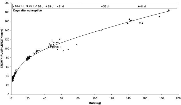
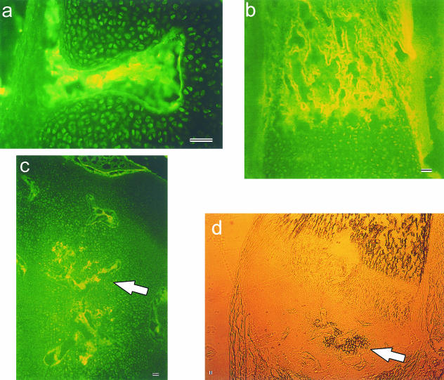
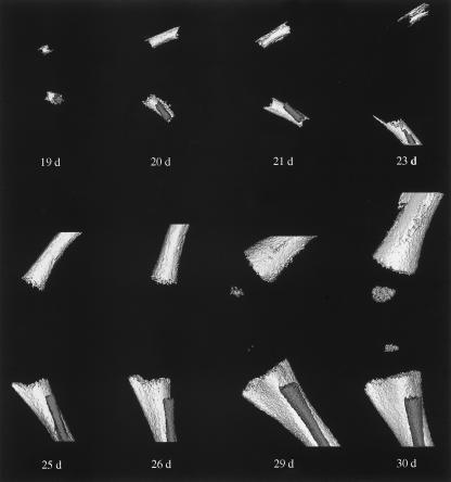
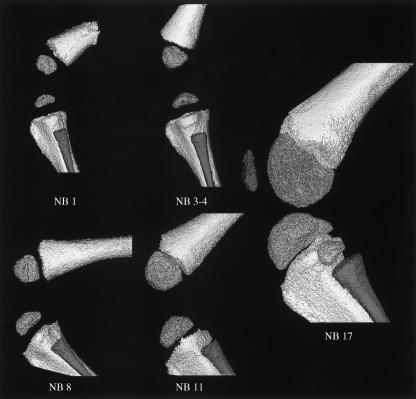
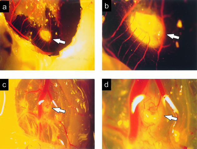
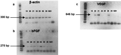
In the developing chondroepiphyses of long bones, the avascular cartilaginous anlage is invaded by numerous blood vessels, through the process of angiogenesis. The objective of this study was to investigate the chronology of this vascular invasion with the spontaneous calcification of the cartilaginous epiphysis during development of the secondary ossification centre in the rabbit distal femur. The time-course of chondroepiphyseal vascular invasion was determined histologically and standardized for eight gestational and four postnatal intervals by plotting kit body mass against crown-rump length. Similarly, microcomputed tomography (micro-CT) helped to visualize calcification at those same gestational and postnatal intervals. To confirm the angiogenic nature of the avascular chondroepiphysis, such samples were assayed on the chick chorio-allantoic membrane (CAM). Neovascular outgrowths from the CAM were apparent 48 h following introduction of an 18-day (gestational) chondroepiphyseal sample. Chondroepiphyseal samples were assayed for the potent developmental angiogenic factors bFGF and VEGF, with the mRNA expression for both these mediators being confirmed using RT-PCR. As angiogenesis and calcification during chondroepiphyseal development occur in a defined tissue environment initially devoid of blood vessels and mineral, those processes provided a unique opportunity to study their progression without complication of injury-related inflammation or extant vasculature and mineral. Furthermore, the discovery of angiogenic, angiostatic or mineral-regulating mediators specific to developing connective tissue may prove useful for analysing the regulation of vascular and mineral pathogenesis in articular tissues.
Figures
Similar articles
-
Matrix metalloproteinase-9 induces the formation of cartilage canals in the chondroepiphysis of the neonatal rabbit.J Bone Joint Surg Am. 2006 Nov;88 Suppl 3:155-61. doi: 10.2106/JBJS.F.00542. J Bone Joint Surg Am. 2006. PMID: 17079382
-
Development of the cartilage canals and the secondary center of ossification in the distal chondroepiphysis of the prenatal human femur.Yale J Biol Med. 1993 May-Jun;66(3):193-202. Yale J Biol Med. 1993. PMID: 8209555 Free PMC article.
-
Angiogenesis in fetal tendon development: spatial and temporal expression of the angiogenic peptide vascular endothelial cell growth factor.Anat Embryol (Berl). 2002 Jul;205(4):263-70. doi: 10.1007/s00429-002-0241-1. Epub 2002 Jun 6. Anat Embryol (Berl). 2002. PMID: 12136256
-
[Angiogenesis, anti-angiogenesis, and tumor suppression].Nihon Yakurigaku Zasshi. 2002 Nov;120(5):285-94. doi: 10.1254/fpj.120.285. Nihon Yakurigaku Zasshi. 2002. PMID: 12491804 Review. Japanese.
-
Therapeutic angiogenesis with vascular endothelial growth factor in peripheral and coronary artery disease: a review.Int J Cardiovasc Intervent. 2003;5(1):27-34. doi: 10.1080/14628840304612. Int J Cardiovasc Intervent. 2003. PMID: 12623562 Review.
Cited by
-
Sulphated glycosaminoglycans and proteoglycans in the developing vertebral column of juvenile Atlantic salmon (Salmo salar).Fish Physiol Biochem. 2015 Aug;41(4):1029-51. doi: 10.1007/s10695-015-0067-4. Epub 2015 May 12. Fish Physiol Biochem. 2015. PMID: 25963942 Free PMC article.
-
The role of cartilage canals in endochondral and perichondral bone formation: are there similarities between these two processes?J Anat. 2005 Apr;206(4):359-72. doi: 10.1111/j.1469-7580.2005.00404.x. J Anat. 2005. PMID: 15817104 Free PMC article.
-
Histomorphometry of Ossification in Functionalised Ceramics with Tripeptide Arg-Gly-Asp (RGD): An In Vivo Study.Life (Basel). 2022 May 20;12(5):761. doi: 10.3390/life12050761. Life (Basel). 2022. PMID: 35629427 Free PMC article.
-
Lentiviral-mediated RNAi knockdown of Cbfa1 gene inhibits endochondral ossification of antler stem cells in micromass culture.PLoS One. 2012;7(10):e47367. doi: 10.1371/journal.pone.0047367. Epub 2012 Oct 9. PLoS One. 2012. PMID: 23056636 Free PMC article.
-
Perlecan immunolocalizes to perichondrial vessels and canals in human fetal cartilaginous primordia in early vascular and matrix remodeling events associated with diarthrodial joint development.J Histochem Cytochem. 2004 Nov;52(11):1405-13. doi: 10.1369/jhc.4A6261.2004. J Histochem Cytochem. 2004. PMID: 15505335 Free PMC article.
References
-
- Ausprunk DH, Folkman J. Migration and proliferation of endothelial cells in preformed and newly formed blood vessels during tumour angiogenesis. Microvascular Res. 1977;14:53–65. - PubMed
-
- Bourin MC. Thrombomodulin: a new proteoglycan. Structure–function relation. Ann. Biol. Clin. 1991;49:199–207. (in French). - PubMed
-
- CCAC (Canadian Council on Animal Care) Guide to the Care and Use of Experimental Animals. Ottawa, Ontario: CCAC; 1994.
-
- Cole AA, Wezeman FH. Perivascular cells in cartilage canals of the developing mouse epiphysis. Am. J. Anat. 1985;174:119–129. - PubMed
Publication types
MeSH terms
Substances
LinkOut - more resources
Full Text Sources
Research Materials
