

Age-related changes in gene expression patterns of matrix metalloproteinases and their collagenous substrates in mandibular condylar cartilage in rats
- PMID: 12924823
- PMCID: PMC1571160
- DOI: 10.1046/j.1469-7580.2003.00196.x
Age-related changes in gene expression patterns of matrix metalloproteinases and their collagenous substrates in mandibular condylar cartilage in rats
Abstract
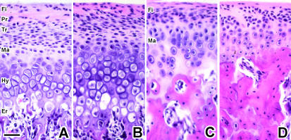
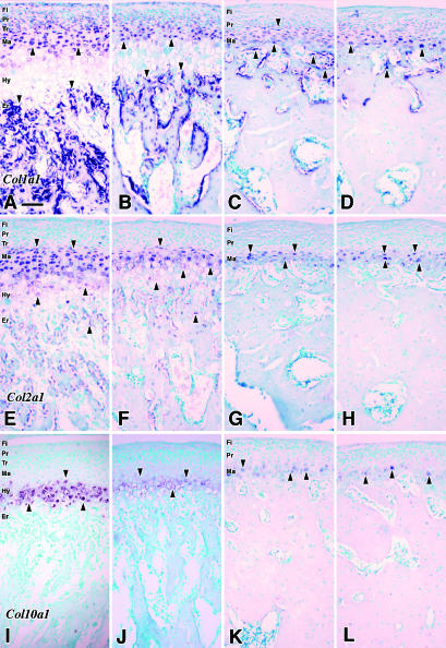
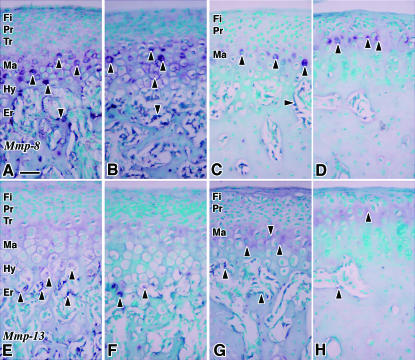
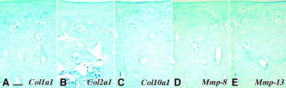
Matrix metalloproteinases (MMPs) have been implicated in physiological cartilage matrix remodelling as well as in pathological and invasive extracellular matrix remodelling of tissue. Age-related changes in the gene expression patterns of MMPs in mandibular condylar cartilages (MCCs) were analysed. We examined the gene expression patterns of Mmp-8 and -13 and their substrates, Col1a1, Col2a1 and Col10a1, in MCC of growing and ageing rats. Temporomandibular joints of male Wistar rats aged 4, 8, 16 and 32 weeks were subjected to in situ hybridization analysis. Histologically, MMCs showed characteristics of growth plate cartilage at ages 4 and 8 weeks, and more closely resembled articular cartilage thereafter. Mmp-8 was expressed in the cells in all cartilaginous cell layers at ages 4 and 8 weeks, and then was localized only in the mature cells at ages 16 and 32 weeks. Whereas Mmp-13 expression was limited to the lowermost hypertrophic chondrocytes in the growth stage, mature chondrocytes instead of hypertrophic chondrocytes expressed Mmp-13 in adult non-hypertrophic MCC. Because Mmp-8 and -13 expression overlapped with Col2a1 and Col10a1, chondrocytes could play a pivotal role in degradation as well as production of the cartilaginous matrix in MCC.
Figures
Similar articles
-
Age-related changes in the expression of gelatinase and tissue inhibitor of metalloproteinase genes in mandibular condylar, growth plate, and articular cartilage in rats.J Mol Histol. 2005 Jun;36(5):355-66. doi: 10.1007/s10735-005-9007-4. Epub 2005 Oct 6. J Mol Histol. 2005. PMID: 16208432
-
Connective tissue growth factor mRNA expression pattern in cartilages is associated with their type I collagen expression.Bone. 2003 Dec;33(6):911-8. doi: 10.1016/j.bone.2003.07.010. Bone. 2003. PMID: 14678850
-
Immunohistochemical localization of matrix metalloproteinase 13 (MMP-13) in mouse mandibular condylar cartilage.J Med Dent Sci. 2003 Sep;50(3):203-11. J Med Dent Sci. 2003. PMID: 15074358
-
Metalloproteinase and tissue inhibitor of metalloproteinase expression in the murine STR/ort model of osteoarthritis.Osteoarthritis Cartilage. 2002 Sep;10(9):722-33. doi: 10.1053/joca.2002.0818. Osteoarthritis Cartilage. 2002. PMID: 12202125
-
[Advances in research of microRNA in the growth and development of mandibular condyle cartilage].Zhonghua Kou Qiang Yi Xue Za Zhi. 2020 Apr 9;55(4):276-279. doi: 10.3760/cma.j.cn112144-20190620-00260. Zhonghua Kou Qiang Yi Xue Za Zhi. 2020. PMID: 32268630 Review. Chinese.
Cited by
-
Evaluation of protein undernourishment on the condylar process of the Wistar rat mandible correlation with insulin receptor expression.J Appl Oral Sci. 2015 Mar-Apr;23(2):135-44. doi: 10.1590/1678-775720140319. J Appl Oral Sci. 2015. PMID: 26018304 Free PMC article.
-
Comparison of age-dependent expression of aggrecan and ADAMTSs in mandibular condylar cartilage, tibial growth plate, and articular cartilage in rats.Histochem Cell Biol. 2006 Sep;126(3):371-80. doi: 10.1007/s00418-006-0171-8. Epub 2006 Apr 1. Histochem Cell Biol. 2006. PMID: 16583222
-
Intermittent Parathyroid Hormone [1-34] Augments Chondrogenesis of the Mandibular Condylar Cartilage of the Temporomandibular Joint.Cartilage. 2021 Oct;12(4):475-483. doi: 10.1177/1947603519833146. Epub 2019 Mar 22. Cartilage. 2021. PMID: 30897936 Free PMC article.
-
Evaluation of the articular cartilage in the knees of rats with induced arthritis treated with curcumin.PLoS One. 2020 Mar 12;15(3):e0230228. doi: 10.1371/journal.pone.0230228. eCollection 2020. PLoS One. 2020. PMID: 32163510 Free PMC article.
-
Impaired extracellular matrix structure resulting from malnutrition in ovariectomized mature rats.Histochem Cell Biol. 2015 Nov;144(5):491-507. doi: 10.1007/s00418-015-1356-9. Epub 2015 Jul 26. Histochem Cell Biol. 2015. PMID: 26210855
References
-
- Armstrong AL, Barrach HJ, Ehrlich MG. Identification of the metalloproteinase stromelysin in the physis. J. Orthop. Res. 2002;20:289–294. - PubMed
-
- Ben Ami Y, von der Mark K, Franzen A, De Bernard B, Lunazzi GC, Silbermann M. Immunohistochemical studies of the extracellular matrix in the condylar cartilage of the human fetal mandible: collagens and noncollagenous proteins. Am. J. Anat. 1991;190:157–166. - PubMed
-
- Buckwalter JA, Rosenberg LC. Electron microscopic studies of cartilage proteoglycans. Electron Microsc. Rev. 1988;1:87–112. - PubMed
-
- Cawston T. Matrix metalloproteinases and TIMPs: properties and implications for the rheumatic diseases. Mol. Med. Today. 1998;4:130–137. - PubMed
-
- Chin JR, Werb Z. Matrix metalloproteinases regulate morphogenesis, migration and remodeling of epithelium, tongue skeletal muscle and cartilage in the mandibular arch. Development. 1997;124:1519–1530. - PubMed
Publication types
MeSH terms
Substances
LinkOut - more resources
Full Text Sources
Medical
Molecular Biology Databases
Miscellaneous
