Case Reports
. 2003 Aug 18;198(4):521-31.
doi: 10.1084/jem.20030701.

Distinct mutations in IRAK-4 confer hyporesponsiveness to lipopolysaccharide and interleukin-1 in a patient with recurrent bacterial infections

Affiliations
Case Reports

Distinct mutations in IRAK-4 confer hyporesponsiveness to lipopolysaccharide and interleukin-1 in a patient with recurrent bacterial infections

Andrei E Medvedev et al. J Exp Med. .

Abstract

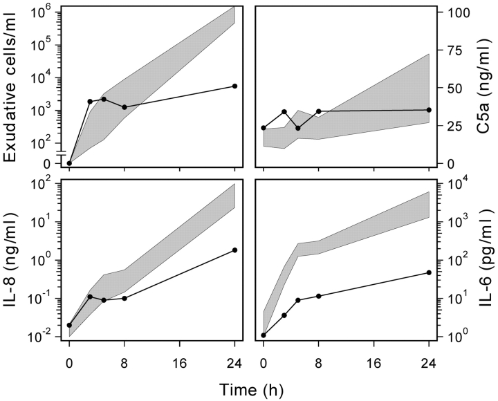
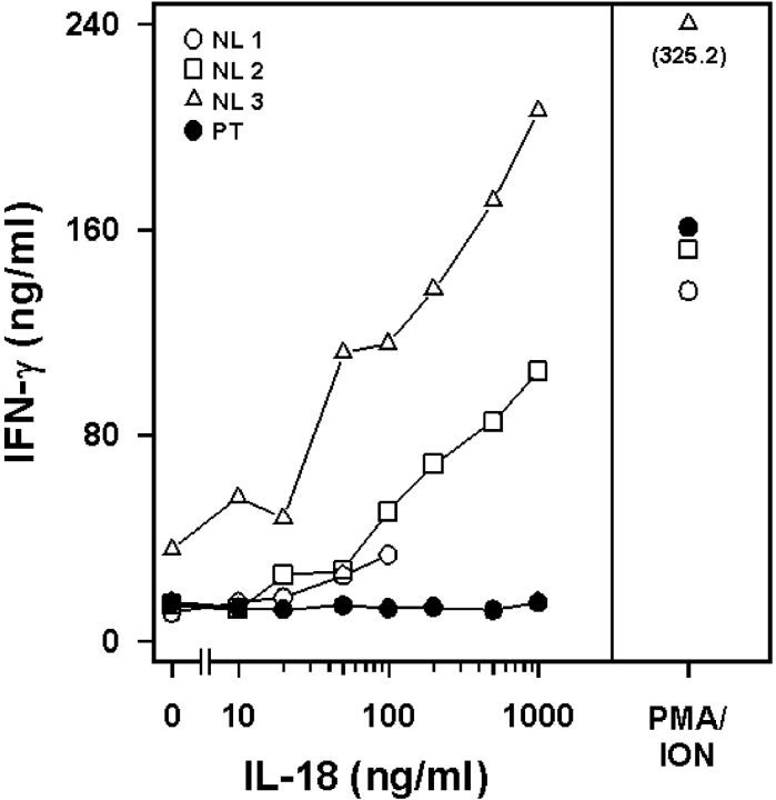
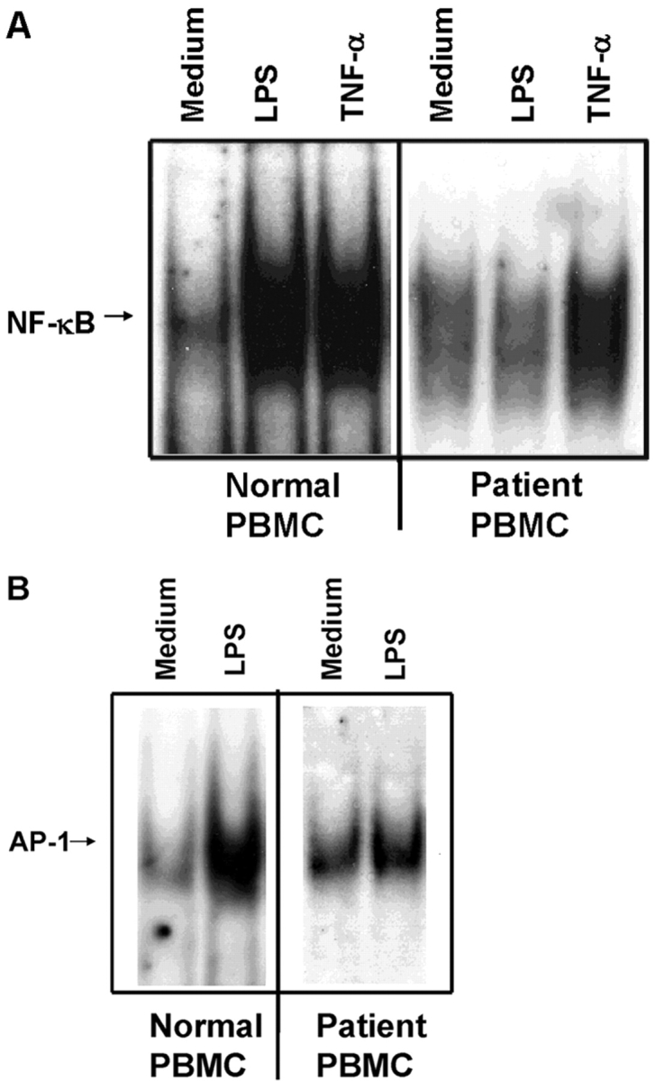
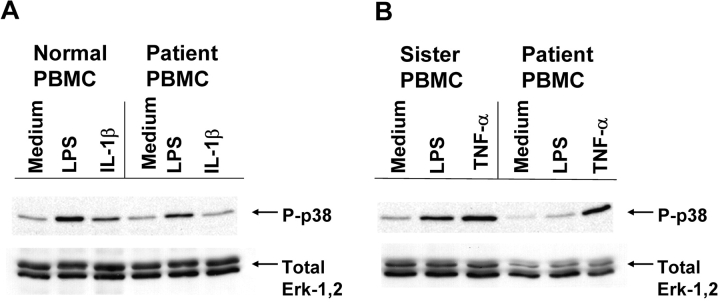
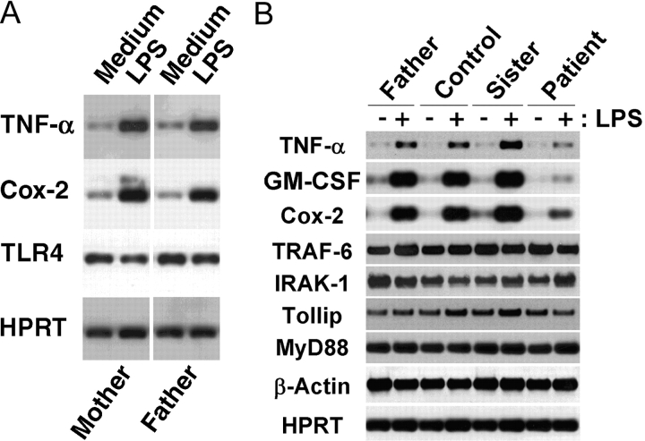
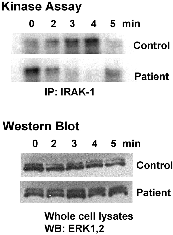
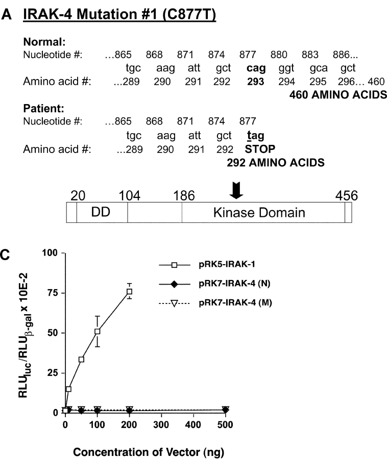
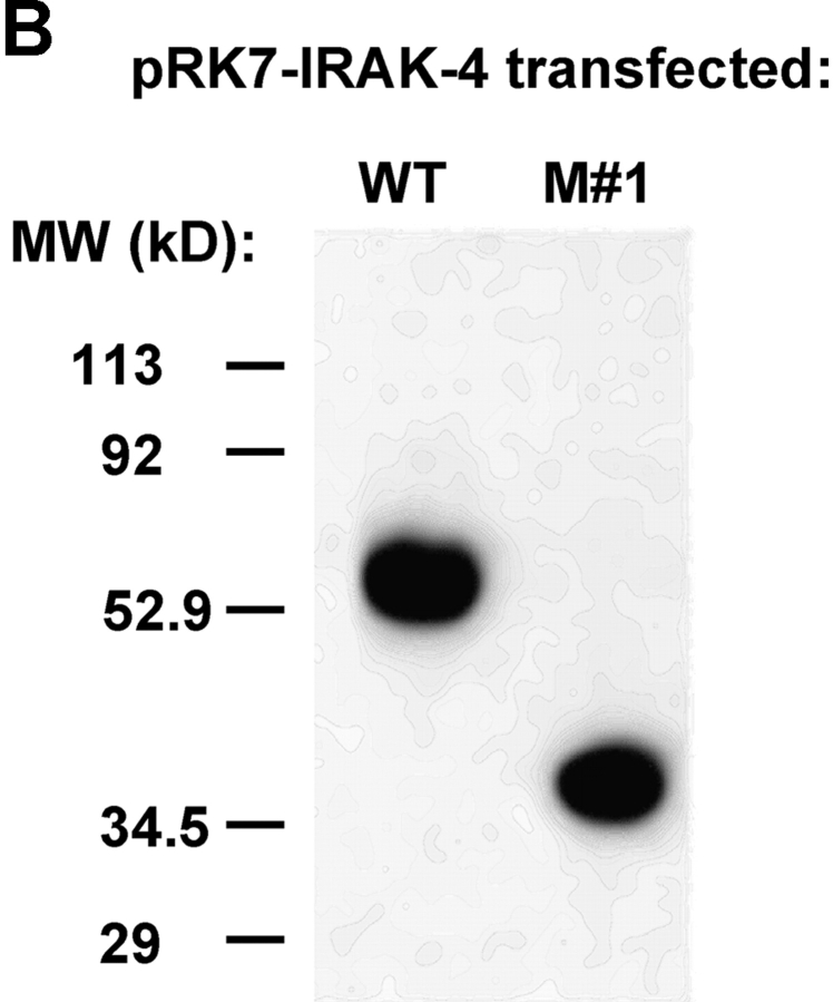
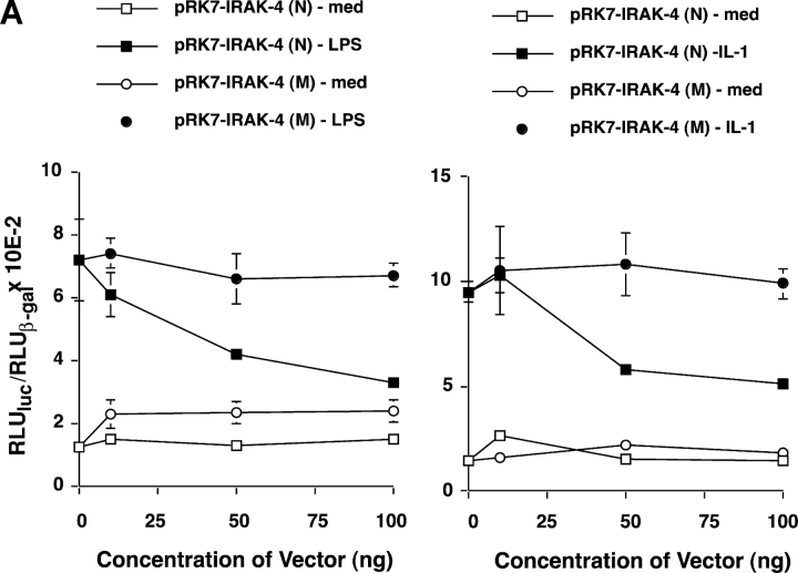
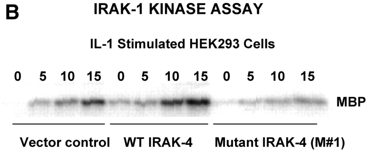
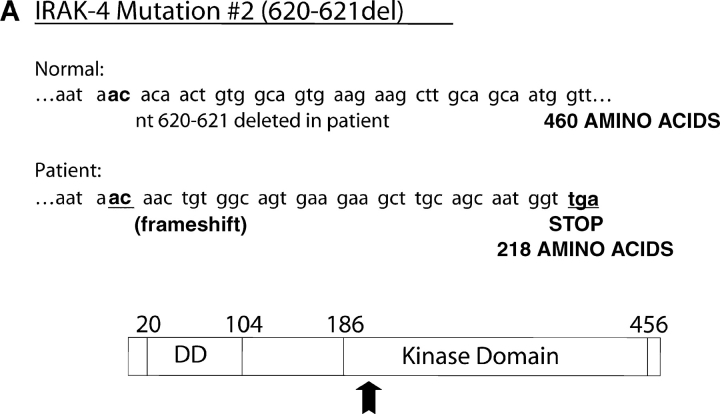
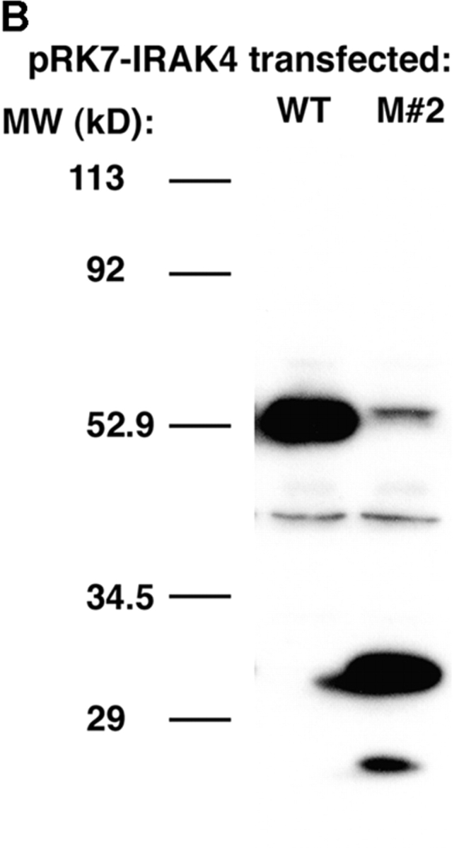
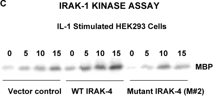
We identified previously a patient with recurrent bacterial infections who failed to respond to gram-negative LPS in vivo, and whose leukocytes were profoundly hyporesponsive to LPS and IL-1 in vitro. We now demonstrate that this patient also exhibits deficient responses in a skin blister model of aseptic inflammation. A lack of IL-18 responsiveness, coupled with diminished LPS and/or IL-1-induced nuclear factor-kappaB and activator protein-1 translocation, p38 phosphorylation, gene expression, and dysregulated IL-1R-associated kinase (IRAK)-1 activity in vitro support the hypothesis that the defect lies within the signaling pathway common to toll-like receptor 4, IL-1R, and IL-18R. This patient expresses a "compound heterozygous" genotype, with a point mutation (C877T in cDNA) and a two-nucleotide, AC deletion (620-621del in cDNA) encoded by distinct alleles of the IRAK-4 gene (GenBank/EMBL/DDBJ accession nos. AF445802 and AY186092). Both mutations encode proteins with an intact death domain, but a truncated kinase domain, thereby precluding expression of full-length IRAK-4 (i.e., a recessive phenotype). When overexpressed in HEK293T cells, neither truncated form augmented endogenous IRAK-1 kinase activity, and both inhibited endogenous IRAK-1 activity modestly. Thus, IRAK-4 is pivotal in the development of a normal inflammatory response initiated by bacterial or nonbacterial insults.


Figures

Figure 1.
Figure 1.
Patient exhibits impaired inflammatory response to blister formation in vivo. The panels show the accumulation of inflammatory cells and mediators in the medium bathing the exposed dermal surface versus time. In each panel, the value at t = 0 represents the background levels found in the 70% autologous serum. The shaded area represents the 95% confidence limits of the arithmetic mean (C5a; linear scale data) and geometric means (exudative cells, IL-8, and IL-6; log scale data) of the responses of normal subjects (n ≥ 7). Patient data (solid line) represents the mean of two experiments.
Figure 2.
Figure 2.
Patient exhibits impaired IL-18 responsiveness in vitro. T cells from normal (NL; open symbols) controls or the patient (PT; closed circles) were stimulated with rIL-18 and levels of IFN-γ measured in culture supernatants. PMA and ionomycin (ION) treatment was used as a positive control for nonreceptor-mediated stimulation of IFN-γ.
Figure 3.
Figure 3.
Patient's PBMCs exhibit impaired NF-κB (A) and AP-1 (B) translocation in response to LPS. PBMCs derived from a control or the patient were stimulated with 1 μg/ml LPS or 100 ng/ml rTNF-α for 60 min, and NF-κB and AP-1 translocation assessed by EMSA.
Figure 4.
Figure 4.
Patient's PBMCs exhibit impaired p38 phosphorylation in response to LPS or IL-1. PBMCs derived from an unrelated control or the patient (A) or the patient's sister and the patient (B) were stimulated with 1 μg/ml LPS, 100 ng/ml rIL-1β, or 100 ng/ml rTNF-α for 15 min. Whole cell lysates were analyzed by Western blot analysis for phosphorylated p38 or total ERK-1,2.
Figure 5.
Figure 5.
Patient's PBMCs exhibit impaired LPS-induced gene expression; LPS-sensitivity of patient's family members is normal. PBMCs were stimulated with 100 ng/ml LPS for 3 h, and RNA was extracted for analysis of gene expression by RT-PCR with Southern blot analysis. (A) Comparison of responses of the patient's mother and father; (B) Comparison of father, sister, unrelated control, and patient responses.
Figure 6.
Figure 6.
Patient's neutrophils exhibit dysregulated IRAK-1 kinase activity. Control and patient neutrophils were stimulated with 100 ng/ml LPS for the indicated times. Cell lysates were immunoprecipitated with anti–IRAK-1 Ab, and immunoprecipitates were assayed for the capacity to phosphorylate MBP. Western blot analysis for total ERK-1,2 was performed on lysates before immunoprecipitation to ensure equal protein concentrations (n = 2).
Figure 7.
Figure 7.
Identification and functionality of a point mutation in patient's IRAK-4. (A) Illustration of C877T substitution mutation (M #1), resulting in a truncated form of IRAK-4. Arrow indicates approximate location of truncation in IRAK-4 protein. (B) Vectors encoding the WT (N) or C877T mutation (M #1) forms of IRAK-4 were expressed in HEK293T cells (5 μg vector/transfection) and cell lysates subjected to Western blot analysis using anti-Flag Ab. (C) Overexpression of WT (N) or C877T (M #1) forms of IRAK-4 in HEK293T cells failed to induce NF-κB–induced reporter activity. Cells were transiently transfected with pELAM-Luc, pCMV-βGal, and the indicated amounts of expression vectors encoding either IRAK-1 or normal (N) and mutated (M) forms of IRAK-4 (total amount of plasmid DNA was kept constant at 1.5 μg per transfection). After recovery for 48 h, NF-κB reporter activity was measured. The data represent the mean ± SEM of a representative experiment (n = 3).
Figure 7.
Figure 7.
Identification and functionality of a point mutation in patient's IRAK-4. (A) Illustration of C877T substitution mutation (M #1), resulting in a truncated form of IRAK-4. Arrow indicates approximate location of truncation in IRAK-4 protein. (B) Vectors encoding the WT (N) or C877T mutation (M #1) forms of IRAK-4 were expressed in HEK293T cells (5 μg vector/transfection) and cell lysates subjected to Western blot analysis using anti-Flag Ab. (C) Overexpression of WT (N) or C877T (M #1) forms of IRAK-4 in HEK293T cells failed to induce NF-κB–induced reporter activity. Cells were transiently transfected with pELAM-Luc, pCMV-βGal, and the indicated amounts of expression vectors encoding either IRAK-1 or normal (N) and mutated (M) forms of IRAK-4 (total amount of plasmid DNA was kept constant at 1.5 μg per transfection). After recovery for 48 h, NF-κB reporter activity was measured. The data represent the mean ± SEM of a representative experiment (n = 3).
Figure 8.
Figure 8.
(A) Overexpression of WT (N), but not C877T mutant (M), IRAK-4 expression vector inhibits LPS- and IL-1–mediated signaling in HEK293T cells. Cells were transiently transfected as described in Fig. 7, permitted to recover for 48 h, and stimulated with medium, LPS, or rIL-1β for 5 h before measurement of NF-κB reporter activity. (B) Overexpression of WT (N), but not C877T mutant (M), IRAK-4 expression vectors differentially modulate IRAK-1 kinase activity. Cells were transiently transfected with control pCDNA3.1, pRK7-IRAK4 (N), or pRK7-IRAK4 (M; 5 μg per transfection). After 40 h recovery, cells were stimulated with rIL-1β for the indicated times, followed by lysis of cells and the IRAK-1 kinase assay (n = 3).
Figure 8.
Figure 8.
(A) Overexpression of WT (N), but not C877T mutant (M), IRAK-4 expression vector inhibits LPS- and IL-1–mediated signaling in HEK293T cells. Cells were transiently transfected as described in Fig. 7, permitted to recover for 48 h, and stimulated with medium, LPS, or rIL-1β for 5 h before measurement of NF-κB reporter activity. (B) Overexpression of WT (N), but not C877T mutant (M), IRAK-4 expression vectors differentially modulate IRAK-1 kinase activity. Cells were transiently transfected with control pCDNA3.1, pRK7-IRAK4 (N), or pRK7-IRAK4 (M; 5 μg per transfection). After 40 h recovery, cells were stimulated with rIL-1β for the indicated times, followed by lysis of cells and the IRAK-1 kinase assay (n = 3).
Figure 9.
Figure 9.
Identification and functionality of deletion mutation in patient's IRAK-4. (A) Illustration of effect of AC deletion at nucleotides 620-621 in the patient (620-621del), resulting in a truncated form of IRAK-4 (M #2). Arrow indicates approximate location of truncation in IRAK-4 protein. (B) Vectors encoding the WT (N) or mutated (M #2) forms of IRAK-4 were expressed in HEK293T cells (10 μg vector/transfection), and cell lysates subjected to Western blot analysis using anti-Flag Ab as described in Fig. 8. (C) Overexpression of WT (N), but not the 620-621 deleted mutant (M #2), IRAK-4 expression vectors (10 μg/transfection) differentially modulate IRAK-1 kinase activity (see Fig. 8 B; n = 3).
Figure 9.
Figure 9.
Identification and functionality of deletion mutation in patient's IRAK-4. (A) Illustration of effect of AC deletion at nucleotides 620-621 in the patient (620-621del), resulting in a truncated form of IRAK-4 (M #2). Arrow indicates approximate location of truncation in IRAK-4 protein. (B) Vectors encoding the WT (N) or mutated (M #2) forms of IRAK-4 were expressed in HEK293T cells (10 μg vector/transfection), and cell lysates subjected to Western blot analysis using anti-Flag Ab as described in Fig. 8. (C) Overexpression of WT (N), but not the 620-621 deleted mutant (M #2), IRAK-4 expression vectors (10 μg/transfection) differentially modulate IRAK-1 kinase activity (see Fig. 8 B; n = 3).

References

    1. Kuhns, D.B., D.A. Long-Priel, and J.I. Gallin. 1997. Endotoxin and IL-1 hyporesponsiveness in a patient with recurrent bacterial infections. J. Immunol. 158:3959–3964. - PubMed
    1. Poltorak, A., X. He, I. Smirnova, M.Y. Liu, C. Van Huffel, X. Du, D. Birdwell, E. Alejos, M. Silva, C. Galanos, et al. 1998. Defective LPS signaling in C3H/HeJ and C57BL/10ScCr mice: mutations in Tlr4 gene. Science. 282:2085–2088. - PubMed
    1. Qureshi, S.T., L. Lariviere, G. Leveque, S. Clermont, K.J. Moore, P. Gros, and D. Malo. 1999. Endotoxin-tolerant mice have mutations in toll-like receptor 4 (Tlr4). J. Exp. Med. 189:615–625. - PMC - PubMed
    1. Medzhitov, R., P. Preston-Hurlburt, E. Kopp, A. Stadleln, C. Chen, S. Ghosh, and C.A. Janeway, Jr. 1998. MyD88 is an adaptor protein in the hToll/IL-1 receptor family signaling pathways. Mol. Cell. 2:253–258. - PubMed
    1. Medzhitov, R.P., and C.A. Janeway, Jr. 2000. Innate immunity. N. Engl. J. Med. 343:338–344. - PubMed

Publication types

MeSH terms

Associated data