. 2003 Aug 18;198(4):615-21.
doi: 10.1084/jem.20030448.

Regulation of dendritic cell migration to the draining lymph node: impact on T lymphocyte traffic and priming

Affiliations

Regulation of dendritic cell migration to the draining lymph node: impact on T lymphocyte traffic and priming

Alfonso MartIn-Fontecha et al. J Exp Med. .

Abstract

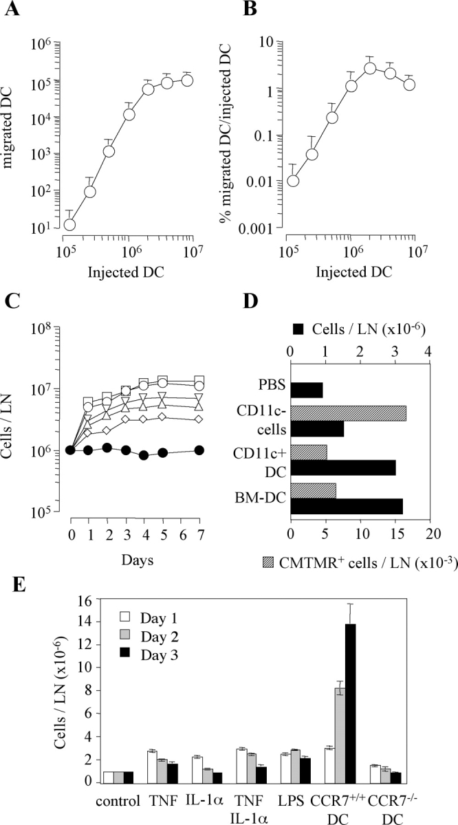
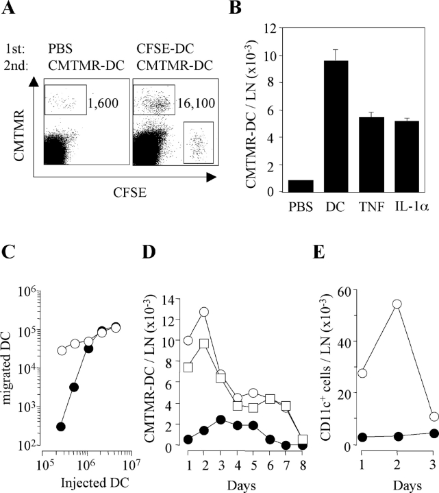
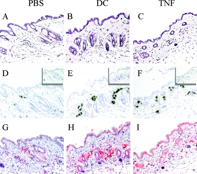
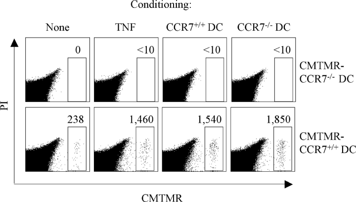
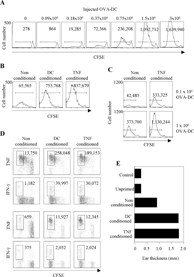
Antigen-pulsed dendritic cells (DCs) are used as natural adjuvants for vaccination, but the factors that influence the efficacy of this treatment are poorly understood. We investigated the parameters that affect the migration of subcutaneously injected mouse-mature DCs to the draining lymph node. We found that the efficiency of DC migration varied with the number of injected DCs and that CCR7+/+ DCs migrating to the draining lymph node, but not CCR7-/- DCs that failed to do so, efficiently induced a rapid increase in lymph node cellularity, which was observed before the onset of T cell proliferation. We also report that DC migration could be increased up to 10-fold by preinjection of inflammatory cytokines that increased the expression of the CCR7 ligand CCL21 in lymphatic endothelial cells. The magnitude and quality of CD4+ T cell response was proportional to the number of antigen-carrying DCs that reached the lymph node and could be boosted up to 40-fold by preinjection of tumor necrosis factor that conditioned the tissue for increased DC migration. These results indicate that DC number and tissue inflammation are critical parameters for DC-based vaccination.


Figures

Figure 1.
Figure 1.
Variable efficiency of DC migration to draining lymph node and consequent lymph node congestion. Increasing numbers of CFSE-labeled mature BM-derived DCs were injected into the footpads of syngeneic mice. The total number of DCs recovered in the draining lymph node (LN; A) and the percentage of injected DCs recovered (B) on day 2 is shown. Mean ± SD of three independent experiments is shown, each performed using groups of two mice per condition. (C) Groups of two mice were injected s.c. with increasing numbers of mature DCs. Total cell number in the pooled draining lymph nodes (LN) was measured at different time points. The number of DCs injected was: 2 × 10⁶ (□), 106 (○), 0.5 × 10⁶ (▿), 0.25 × 10⁶ (▵), and 0.125 × 10⁶ (⋄). Mice injected with PBS were used as control (•). The experiment was repeated with comparable results. (D) Mice were injected s.c. with PBS, 106 BM-derived DC (BM-DC), 106 CD11c+ DCs isolated from the spleen or 10 × 106 CD11c spleen cells that had been labeled with CMTMR. The total cell number in the lymph node (solid bars) and the number of CMTMR+ migrated cells (shaded bars) are shown. One out of three experiments performed is shown. (E) Groups of three mice per condition per time point were injected s.c. with PBS (control), 300 ng TNF, and/or IL-1α, 100 μg LPS, or 106 BM-derived syngeneic DCs from CCR7+/+ or CCR7−/− mice. The total cell number (means ± SD) in the draining lymph node was measured on days 1, 2, and 3.
Figure 2.
Figure 2.
The efficiency of DC migration to draining lymph node is increased by TNF, IL-1, and by DCs themselves. (A) PBS or 106 CFSE-labeled DCs were injected into the footpads of syngeneic mice. 24 h later, the mice received a second injection of 106 CMTMR-labeled DCs in the same footpad. Mice were killed 24 h after the second injection and the number of CMTMR+ DCs migrated to the draining lymph nodes was analyzed. (B) The experiment was performed as in A after the injection of PBS, unlabeled 106 DCs, TNF, or IL-1α. Mean of three experiments each performed with groups of two mice per condition is shown. (C) Footpads pretreated with either PBS (•) or TNF (○) were injected with increasing doses of CMTMR-labeled DCs and the number of migrated DCs recovered in the draining lymph node was measured 2 d later. (D) Footpads of mice pretreated with PBS (•), TNF (□), or 106 unlabeled DCs (○) were injected with 106 CMTMR DCs and the number of migrated DCs was evaluated daily. Values are from pooled lymph nodes of two mice per time point. (E) CMFDA was applied to the skin of mice that had been pretreated with either PBS (•) or TNF (○). The number of fluorescent endogenous CD11c+ DCs recovered in the draining lymph node is shown.
Figure 3.
Figure 3.
CCL21 is up-regulated on lymphatic endothelial cells after injection of mature DCs or TNF. Mice were injected intradermally with PBS, 105 syngeneic DCs, or 10 ng TNF. 8 h later, skin samples were collected and embedded in paraffin. Serial sections were stained with hematoxylin and eosin (A–C), or hybridized with antisense (D–F) or sense (inset) 35S-labeled CCL21 riboprobe or stained with a polyclonal antibody to mouse CCL21 (G–I). ×20.
Figure 4.
Figure 4.
CCR7−/− DCs fail to migrate to the draining lymph node but efficiently condition the tissue for increased migration of CCR7+/+ DCs. Mice (two per condition) were injected with 106 CCR7+/+ or CCR7−/− syngeneic DCs, TNF, or PBS. 24 h later, all mice received a second injection at the same site of 106 CMTMR-labeled CCR7−/− or CCR7+/+ DCs. The total number of migrated CMTMR-labeled DCs was measured in the draining lymph node after an additional 24 h (gate). Live cells gated as propidium iodide (PI) negative are shown. One of two experiments performed is shown.
Figure 5.
Figure 5.
Impact of DC migration and tissue conditioning on T cell response. (A) 3 × 10⁶ KJ1–26+ T cells from DO11.10 mice were labeled with CFSE and adoptively transferred into syngeneic BALB/c mice. Control or conditioned mice were primed by injecting OVA323–339–pulsed syngeneic DCs in the footpad. CFSE profiles (day 3) of KJ1.26-gated cells and the absolute number of proliferating T cells after injection of increasing numbers of OVA-pulsed DCs. One representative experiment out of three is shown. (B) CFSE profiles (day 4) of KJ1-26–gated cells and total number of proliferating cells in mice conditioned with PBS, TNF, or 106 syngeneic DCs and primed by 105 OVA-pulsed DCs. Data are from pooled lymph nodes from two mice. (C) CFSE profile (day 4) of KJ1-26–gated T cells and total number of proliferating T cells in control or TNF-conditioned mice primed by 0.1 × 10⁶ or 106 OVA-pulsed CMTMR-labeled DCs. The number of DCs recovered in the same lymph nodes was as follows: nonconditioned, 0.1 × 10⁶ DCs injected, <10; TNF-conditioned, 0.1 × 10⁶ DCs injected, 1,180; nonconditioned, 106 DCs injected, 1,600; TNF-conditioned, 106 DCs injected, 9,320. (D) TNF and IFN-γ production elicited by PMA and ionomycin in KJ1-26–gated cells recovered from draining (top panels) and nondraining (bottom panels) lymph nodes. The total number of cytokine-secreting cells was calculated from the percentage and the total number of cells and is indicated in the dot plot. (E) Mice were challenged by injection of 10 μg OVA in the ear pinna 7 d after priming. Controls include naive mice and mice adoptively transferred with naive DO11.10 T cells (unprimed). Ear thickness was measured 24 h after challenge. One out of two experiments performed is shown.

References

    1. Banchereau, J., and R.M. Steinman. 1998. Dendritic cells and the control of immunity. Nature. 392:245–252. - PubMed
    1. Dieu, M.C., B. Vanbervliet, A. Vicari, J.M. Bridon, E. Oldham, S. Ait-Yahia, F. Briere, A. Zlotnik, S. Lebecque, and C. Caux. 1998. Selective recruitment of immature and mature dendritic cells by distinct chemokines expressed in different anatomic sites. J. Exp. Med. 188:373–386. - PMC - PubMed
    1. Sallusto, F., P. Schaerli, P. Loetscher, C. Schaniel, D. Lenig, C.R. Mackay, S. Qin, and A. Lanzavecchia. 1998. Rapid and coordinated switch in chemokine receptor expression during dendritic cell maturation. Eur. J. Immunol. 28:2760–2769. - PubMed
    1. Ngo, V.N., H.L. Tang, and J.G. Cyster. 1998. Epstein-Barr virus–induced molecule 1 ligand chemokine is expressed by dendritic cells in lymphoid tissues and strongly attracts naive T cells and activated B cells. J. Exp. Med. 188:181–191. - PMC - PubMed
    1. Gunn, M.D., K. Tangemann, C. Tam, J.G. Cyster, S.D. Rosen, and L.T. Williams. 1998. A chemokine expressed in lymphoid high endothelial venules promotes the adhesion and chemotaxis of naive T lymphocytes. Proc. Natl. Acad. Sci. USA. 95:258–263. - PMC - PubMed

Publication types