

Review
doi: 10.1172/JCI19552.
The potential for gene repair via triple helix formation
Affiliations
- PMID: 12925687
- PMCID: PMC171401
- DOI: 10.1172/JCI19552
Item in Clipboard
Review
The potential for gene repair via triple helix formation
J Clin Invest.
2003 Aug.
Abstract
Triplex-forming oligonucleotides (TFOs) can bind to polypurine/polypyrimidine regions in DNA in a sequence-specific manner. The specificity of this binding raises the possibility of using triplex formation for directed genome modification, with the ultimate goal of repairing genetic defects in human cells. Several studies have demonstrated that treatment of mammalian cells with TFOs can provoke DNA repair and recombination, in a manner that can be exploited to introduce desired sequence changes. This review will summarize recent advances in this field while also highlighting major obstacles that remain to be overcome before the application of triplex technology to therapeutic gene repair can be achieved.
Figures

Diagrammatic depiction of a DNA triple helix, with the third strand binding in the major groove.
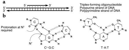
Diagram of the pyrimidine motif for triple helix formation. (a) Orientation of the third strand in the pyrimidine triple-helix motif. Note that the third strand is parallel in terms of 5′ to 3′ orientation with respect to the purine-rich strand of the duplex target. (b) Base triplets formed in the pyrimidine motif and illustration of the Hoogsteen hydrogen bonds that stabilize triple-helix formation.

Diagram of the purine motif for triple-helix formation. (a) Orientation of the third strand in the purine triple-helix motif. Note that the third strand is oriented antiparallel in terms of 5′ to 3′ direction with respect to the purine-rich strand of the duplex target. (b) Base triplets formed in the purine motif and illustration of the reverse Hoogsteen hydrogen bonds that stabilize triple-helix formation.
References
-
- Felsenfeld G, Davies DR, Rich A. Formation of a three stranded polynucleotide molecule. J. Am. Chem. Soc. 1957;79:2023–2024.
-
- Thuong NT, Helene C. Sequence specific recognition and modification of double helical DNA by oligonucleotides. Angewandte Chemie. Intl. Ed. Eng. 1993;32:666–690.
-
- Frank-Kamenetskii MD, Mirkin SM. Triplex DNA structures. Annu. Rev. Biochem. 1995;64:65–95. - PubMed
-
- Manor H, Rao BS, Martin RG. Abundance and degree of dispersion of genomic d(GA)n.d(TC)n sequences. J. Mol. Evol. 1988;27:96–101. - PubMed
Publication types
MeSH terms
Substances
Grants and funding
LinkOut - more resources
Full Text Sources
Other Literature Sources
Medical
