. 2003 Aug;112(4):503-16.
doi: 10.1172/JCI17913.

Role for integrin-linked kinase in mediating tubular epithelial to mesenchymal transition and renal interstitial fibrogenesis

Affiliations

Role for integrin-linked kinase in mediating tubular epithelial to mesenchymal transition and renal interstitial fibrogenesis

Yingjian Li et al. J Clin Invest. 2003 Aug.

Erratum in

  • J Clin Invest. 2004 Feb;113(3):491

Abstract

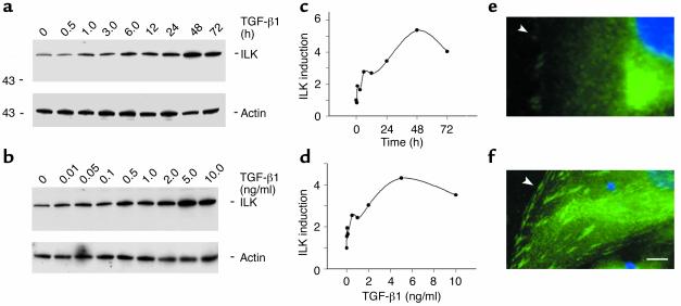
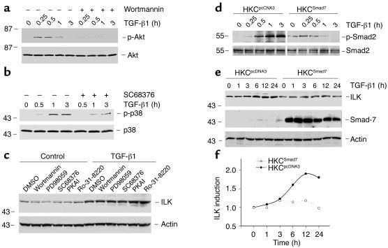
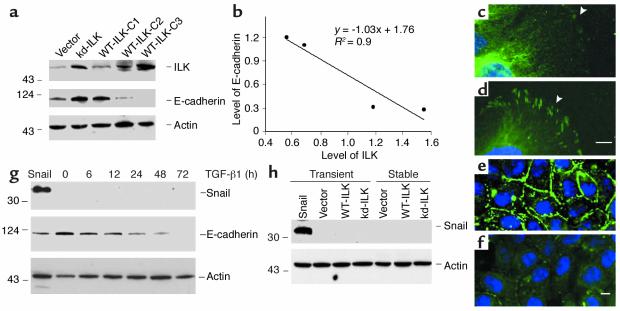
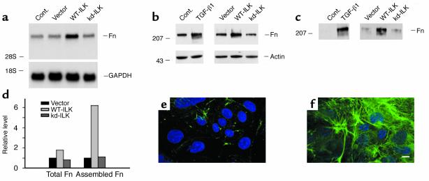
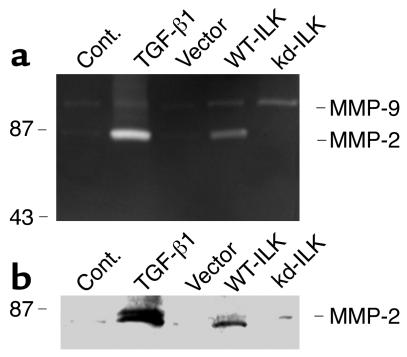
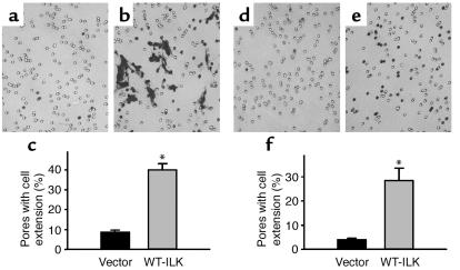
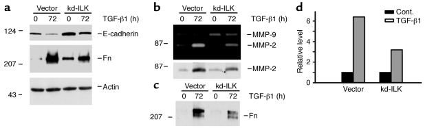
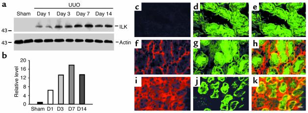
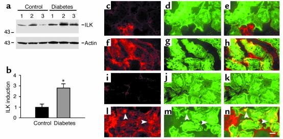
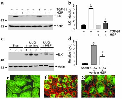
Under pathologic conditions, renal tubular epithelial cells can undergo epithelial to mesenchymal transition (EMT), a phenotypic conversion that is believed to play a critical role in renal interstitial fibrogenesis. However, the underlying mechanism that governs this process remains largely unknown. Here we demonstrate that integrin-linked kinase (ILK) plays an important role in mediating tubular EMT induced by TGF-beta1. TGF-beta1 induced ILK expression in renal tubular epithelial cells in a time- and dose-dependent manner, which was dependent on intracellular Smad signaling. Forced expression of ILK in human kidney proximal tubular epithelial cells suppressed E-cadherin expression and induced fibronectin expression and its extracellular assembly. ILK also induced MMP-2 expression and promoted cell migration and invasion in Matrigel. Conversely, ectopic expression of a dominant-negative, kinase-dead form of ILK largely abrogated TGF-beta1-initiated tubular cell phenotypic conversion. In vivo, ILK was markedly induced in renal tubular epithelia in mouse models of chronic renal diseases, and such induction was spatially and temporally correlated with tubular EMT. Moreover, inhibition of ILK expression by HGF was associated with blockade of tubular EMT and attenuation of renal fibrosis. These findings suggest that ILK is a critical mediator for tubular EMT and likely plays a crucial role in the pathogenesis of chronic renal fibrosis.


Figures

Figure 1
Figure 1
TGF-β1 induces ILK expression in renal tubular epithelial cells. (ad) Western blot analyses show that TGF-β1 induced ILK protein expression in a time- and dose-dependent manner. HKC cells were incubated with either the same concentration of TGF-β1 (2 ng/ml) for various periods of time (a and c) or with increasing amounts of TGF-β1 for 24 hours (b and d). Cell lysates were immunoblotted with Ab’s against ILK and actin, respectively. (c and d) Graphic presentation of relative ILK abundance (fold induction) normalized to actin. (e and f) Immunofluorescence staining shows the localization of ILK in control (e) or TGF-β1–treated HKC cells (f). Arrowheads indicate positive ILK staining. Scale bar, 5 μm.
Figure 2
Figure 2
ILK induction by TGF-β1 in renal epithelial cells is dependent on Smad signaling. (ac) Pharmacological inhibition of different signal transduction pathways does not affect ILK induction by TGF-β1. HKC cells were pretreated with either various chemical inhibitors or vehicle (DMSO) for 30 minutes, followed by incubating in the absence or presence of 2 ng/ml TGF-β1 for 0.25, 0.5, 1 hour, 3 hours (a and b) and for 24 hours (c), respectively. Specific inhibitors for PI3K (10 nM wortmannin), Mek1 (10 μM PD98059), p38 MAPK (20 μM SC68376), PKA (0.3 μM PKA inhibitor [PKAI]), and PKC (50 nM Ro-31-8220) were used, respectively. Cell lysates were immunoblotted with Ab’s against phosphospecific Akt (p-Akt) and total Akt (a), phosphospecific and total p38 MAPK (p-p38 and p38, respectively) (b), ILK, and actin (c), respectively. (df) Overexpression of inhibitory Smad-7 abolishes ILK induction by TGF-β1. A stable cell line overexpressing inhibitory Smad-7 (HKCSmad7) was established by transfection of Smad-7 expression vector. A cell line with mock transfection of empty pcDNA3 vector (HKCpcDNA3) was used as control. Cells were treated with 2 ng/ml of TGF-β1 for various periods of time as indicated. (d) Cell lysates were blotted with phosphospecific (p-Smad2) and total Smad-2, respectively. (e) Cell lysates were blotted with Ab’s against ILK, Smad-7, and actin, respectively. (f) Graphical presentation of relative ILK abundance normalized to actin following TGF-β1 treatment in HKCpcDNA3 and HKCSmad7 cells. AU, arbitrary units.
Figure 3
Figure 3
Forced expression of ILK suppresses E-cadherin expression in tubular epithelial cells in a dose-dependent manner. Stable cell lines were established by transfection of WT-ILK and kd-ILK expression vectors. A cell line with mock transfection of empty vector pUSEamp plasmid (Vector) was used as control. (a) Cell lysates were immunoblotted with Ab’s against ILK, E-cadherin, and actin, respectively. Numbers (C1, C2, and C3) indicate three individual clones that express different levels of ILK. (b) Linear regression shows a close association between ILK and E-cadherin in tubular epithelial cells. The correlation coefficients (R2) are shown. The relative abundances of ILK and E-cadherin were normalized to actin. (cf) Immunofluorescence staining shows an inverse relationship between ILK and E-cadherin expression in HKC cells transfected with empty vector (c and e) or WT-ILK (d and f). (c and d) ILK staining. (e and f) E-cadherin staining. Scale bars, 5 μm. (g and h) Suppression of E-cadherin expression by ILK is independent of Snail. Neither TGF-β1 (g) nor ILK (h) induced Snail expression in tubular epithelial cells. HKC cells were treated with 2 ng/ml of TGF-β1 for various periods of time as indicated (g), or transfected (either transiently or stably) with WT-ILK and kd-ILK expression vectors (h). HKC cells transiently transfected with Snail expression vector served as positive control for Snail protein expression.
Figure 4
Figure 4
Forced expression of ILK induces fibronectin expression and its extracellular assembly. (a) Northern blot analysis shows that forced expression of ILK induces fibronectin (Fn) mRNA expression in tubular epithelial cells. Total RNA was isolated from different cell lines as indicated and hybridized with cDNA probes of fibronectin and GAPDH, respectively. Cont., control parental HKC cells; vector, HKC cells transplanted with empty vector. (b) Western blot demonstrates that either treatment with TGF-β1 or forced expression of ILK induced total fibronectin protein expression in tubular epithelial cells. Cell lysates were immunoblotted with Ab’s against fibronectin and actin, respectively. (c) Expression of ILK promotes the extracellular assembly of fibronectin. Extracellular protein extracts were prepared from various cells as indicated and subjected to Western blot analyses using fibronectin Ab. (d) Graphical presentation shows the effects of ILK expression on the relative extent of total fibronectin expression and its extracellular assembly. Data are presented as fold induction over the empty vector control cells. (e and f) Immunofluorescence staining demonstrates the extracellular assembly of fibronectin in tubular epithelial cells transfected with empty vector (e) or WT-ILK (f). Scale bar, 5 μm.
Figure 5
Figure 5
Expression of ILK induces MMP-2 expression and secretion by tubular epithelial cells. (a) Zymographic analysis shows the proteolytic activity of MMP-2 and MMP-9 in the supernatants of different cell lines. The locations of MMP-9 and MMP-2 are indicated. The samples from HKC cells treated without or with 2 ng/ml of TGF-β1 were loaded in the adjacent lanes to serve as controls for MMP-2 induction. (b) Western blot analysis demonstrates that expression of ILK induces MMP-2 expression by tubular epithelial cells. Equal amounts of the supernatants derived from the HKC lines with overexpression of either WT-ILK, kd-ILK, or empty vector were immunoblotted with specific Ab against MMP-2.
Figure 6
Figure 6
Forced expression of ILK enhances the migration and invasion capacity of tubular epithelial cells. (ac) Boyden chamber motility assay demonstrates enhanced cell migration in tubular epithelial cells overexpressing ILK. Control (vector) (a) and ILK-overexpressing WT-ILK-C3 (b) cells were seeded on Transwell membranes and incubated for 5 days. Cells and cell extensions that migrated through the pores of Transwell plates were counted and reported (c). *P < 0.01, n = 3. (df) Matrigel invasion assay shows an enhanced invasion capacity of the tubular epithelial cells overexpressing ILK. Control (empty vector) (d) and ILK-overexpressing WT-ILK-C3 (e) cells were seeded on top of Matrigel in the Transwell filters of the Boyden chamber and incubated for 5 days. Cell extensions that invaded the Matrigel and migrated through the pores of the Transwell plates were counted and reported (f). *P < 0.01, n = 3.
Figure 7
Figure 7
Ectopic expression of kd-ILK largely blocks TGF-β1–induced phenotypic transition of tubular epithelial cells. (a) HKC cells transfected with empty vector or kd-ILK were incubated with or without 2 ng/ml of TGF-β1 for 72 hours. Western blot analyses demonstrate that ectopic expression of kd-ILK largely blocked TGF-β1–initiated E-cadherin suppression and fibronectin induction in tubular epithelial cells. (b) Zymographic analysis (upper panel) and Western blot (lower panel) show that expression of kd-ILK reduces MMP-2 expression induced by TGF-β1. (c and d) Expression of kd-ILK attenuates the extracellular assembly of fibronectin after TGF-β1 treatment. Extracellularly assembled fibronectin was analyzed by Western blot analyses (c). Graphical presentation (d) shows the effect of kd-ILK expression on the relative abundance of assembled fibronectin induced by TGF-β1. Data are presented as fold induction over cells not treated with TGF-β1.
Figure 8
Figure 8
Induction of ILK expression occurs specifically in renal epithelia in obstructive nephropathy. (a and b) Western blot analysis shows marked induction of ILK in the fibrotic kidney induced by UUO in a time-dependent manner. Kidney homogenates were immunoblotted with Ab’s against ILK and actin, respectively. The relative abundances of ILK are presented in b after normalization with actin. Samples from two individual animals were used at each timepoint. (ck) Immunofluorescence staining shows localization of ILK (red) and the tubular cell marker lectin (green) in the sham kidney (ce) and obstructed kidneys after 3 (fh) and 7 days (ik), respectively. Scale bar, 20 μm.
Figure 9
Figure 9
Induction of ILK expression in renal epithelia in diabetic nephropathy is associated with tubular EMT. (a and b) Western blot analysis shows induction of ILK expression in diabetic nephropathy in mice. Whole-tissue homogenates derived from control or diabetic mice were immunoblotted with Ab’s against ILK and actin, respectively. The relative abundance of ILK (fold induction) is presented in b after normalization with actin. *P < 0.05 (n = 3). (ch) Immunofluorescence staining shows localization of ILK (red) and the tubular cell marker lectin (green) in control (ce) and diabetic kidneys (fh), respectively. (in) Immunofluorescence staining demonstrates tubular EMT in diabetic nephropathy, as illustrated by colocalization of α-smooth muscle actin (red) and lectin (green) in diabetic kidneys (ln) (arrowheads). (ik) Control kidney. Scale bar, 20 μm.
Figure 10
Figure 10
Inhibition of ILK expression in tubular epithelial cells by HGF in vitro and in vivo. (a and b) HGF inhibits ILK expression induced by TGF-β1 in tubular epithelial cells in vitro. HKC cells were treated with TGF-β1 (2 ng/ml), HGF (40 ng/ml), or both for 48 hours. Representative Western blot (a) and graphical presentation (b) show the relative abundances of ILK (fold induction) after various treatments. Data are presented as mean ± SEM of three independent experiments. *P < 0.01 vs. normal control; P < 0.01 vs. TGF-β1 alone. (c and d) HGF inhibits renal ILK expression in obstructive nephropathy. Whole-tissue homogenates derived from the kidneys at 7 days after surgery were immunoblotted with Ab’s against ILK and actin, respectively. The relative abundances of ILK (fold induction) are presented in d after normalization with actin. *P < 0.01 vs. sham; P < 0.01 vs. vehicle control (n = 3). (eg) Double immunofluorescence staining shows localization of ILK (red) and the tubular cell marker lectin (green) in the kidneys at 7 days after surgery in the sham (e), UUO with vehicle (f), and UUO with HGF (g) groups, respectively. Scale bar, 20 μm. Inhibition of ILK expression by HGF is accompanied by blockage of tubular EMT and attenuation of renal fibrosis, as previously reported (6).

Similar articles

Cited by

References

    1. Strutz F, Muller GA. Transdifferentiation comes of age. Nephrol. Dial. Transplant. 2000;15:1729–1731. - PubMed
    1. Yang J, Liu Y. Dissection of key events in tubular epithelial to myofibroblast transition and its implications in renal interstitial fibrosis. Am. J. Pathol. 2001;159:1465–1475. - PMC - PubMed
    1. Iwano M, et al. Evidence that fibroblasts derive from epithelium during tissue fibrosis. J. Clin. Invest. 2002;110:341–350. doi:10.1172/JCI200215518. - PMC - PubMed
    1. Yang J, et al. Disruption of tissue-type plasminogen activator gene in mice reduces renal interstitial fibrosis in obstructive nephropathy. J. Clin. Invest. 2002;110:1525–1538. doi:10.1172/JCI200216219. - PMC - PubMed
    1. Zeisberg M, et al. Renal fibrosis: collagen composition and assembly regulates epithelial-mesenchymal transdifferentiation. Am. J. Pathol. 2001;159:1313–1321. - PMC - PubMed

Publication types

MeSH terms

Substances