. 2003 Aug;112(4):517-26.
doi: 10.1172/JCI18028.

Cathepsin V is involved in the degradation of invariant chain in human thymus and is overexpressed in myasthenia gravis

Affiliations

Cathepsin V is involved in the degradation of invariant chain in human thymus and is overexpressed in myasthenia gravis

Eva Tolosa et al. J Clin Invest. 2003 Aug.

Abstract

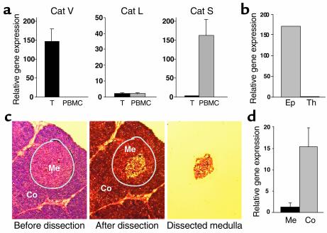
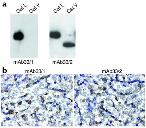
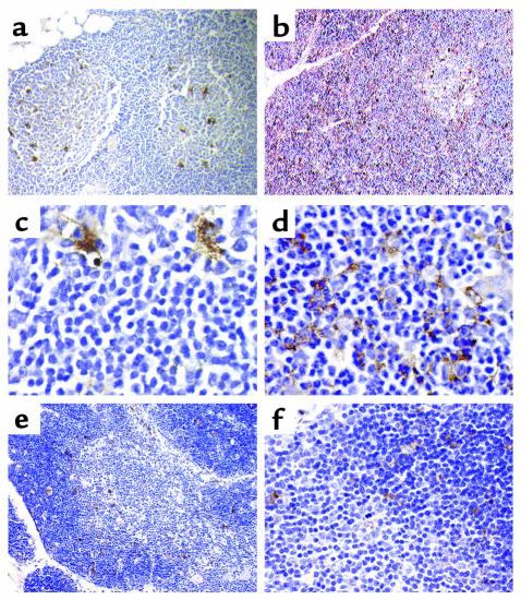
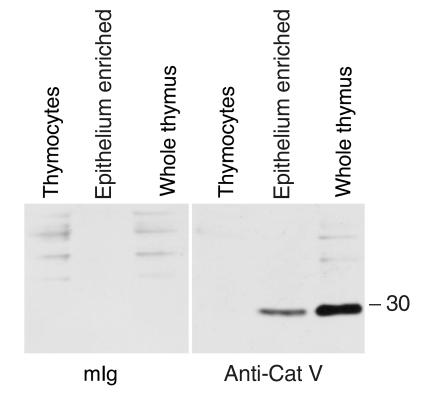
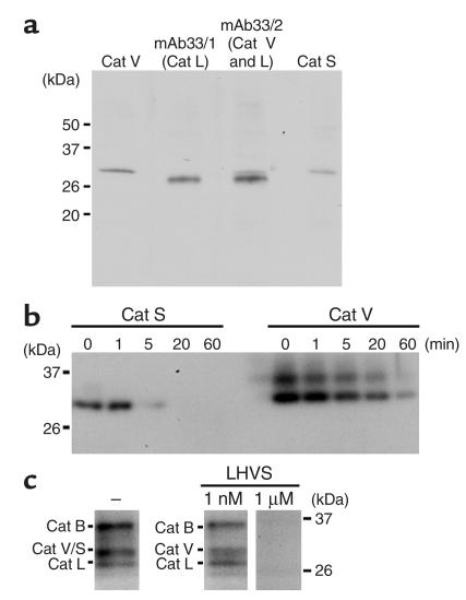
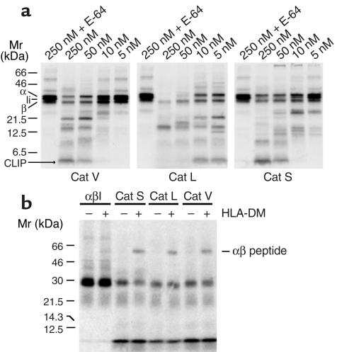
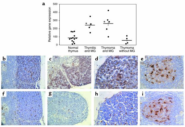
Stepwise degradation of the invariant chain (Ii) is required for the binding of antigenic peptides to MHC class II molecules. Cathepsin (Cat) L in the murine thymus and Cat S in peripheral APCs have both been implicated in the last step of Ii degradation that gives rise to the class II-associated invariant chain peptides (CLIP). Cat V has been recently described as highly homologous to Cat L and exclusively expressed in human thymus and testis, but with no mouse orthologue. We report that Cat V is the dominant cysteine protease in cortical human thymic epithelial cells, while Cat L and Cat S seem to be restricted to dendritic and macrophage-like cells. Active Cat V in thymic lysosomal preparations was demonstrated by active-site labeling. Recombinant Cat V was capable of converting Ii into CLIP efficiently, suggesting that Cat V is the protease that controls the generation of alphabeta-CLIP complexes in the human thymus, in analogy to Cat L in mouse. Comparison of Cat V expression between thymi from patients with myasthenia gravis and healthy controls revealed a significantly higher expression level in the pathological samples, suggesting a potential involvement of this protease in the immunopathogenesis of myasthenia gravis, an autoimmune disease almost invariably associated with thymic pathology.


Figures

Figure 1
Figure 1
mRNA expression of Cats V, L, and S. (a) Real-time RT-PCR analysis for the expression of Cats V, L, and S in thymus (T, black bars) and PBMCs (gray bars). The graphs display the averaged values of seven thymi and five PBMC samples. (b) Cat V in the thymus is exclusively found in an epithelial cell-enriched fraction (Ep) and could not be detected in thymocytes (Th). (c and d) Laser capture microdissection of the thymic medulla (Me, black bar) and cortex (Co, gray bar), followed by RT-PCR analysis shows that Cat V is mostly found in the cortex of the thymus.
Figure 2
Figure 2
Specificity of mAb33/1 and mAb33/2 for human Cats V and L. (a) Western blot analysis of recombinant human Cats V and L using mAb33/1 and mAb33/2. Both enzymes, 0.05 μg, were loaded onto 12% SDS Tris-glycine gels and antibody dilutions of 1:5000 were used. The mAb33/2 cross reacts with both proteases, whereas mAb33/1 only recognizes Cat L. (b) The mAb33/1 and mAb33/2 recognize Cat L in human spleen tissue sections and are indistinguishable in their sensitivity toward Cat L antigen in paraffin-embedded tissue section. The ABC staining kit, Super Sensitive Ready-To-Use Detection System (Biogenex), was used for immunohistochemical protein localization.
Figure 3
Figure 3
Immunohistochemical analysis of Cats V, L, and S expression in thymus. Mouse monoclonal antihuman Cat L antibodies mAb33/1 (a and c; recognizes only Cat L), mAb33/2 (b and d; cross reacts with human Cats V and L) and a rabbit polyclonal antibody against Cat S (e and f) were used to localize these proteases in human thymus. a and c show the expression of Cat L in macrophage-like cells of the normal thymus. b shows the expression of Cat V in thymic cortex. Immunopositive cells in the medulla may express either Cats L and V or only Cat L. d demonstrates the expression in cortical thymic epithelial cells, which do not express Cat L (see c). e and f show the discrete staining for Cat S both in the cortex and medulla with a rabbit polyclonal antibody specific for Cat S. All magnifications are ×1000 with the exception of a and e (×200) and b (×400). Tissue sections were counterstained with hematoxylin.
Figure 4
Figure 4
Western blot. Purified thymocytes, an epithelium-enriched fraction of human thymus and whole thymus, were analyzed by Western blot with anti-Cat V-specific mAb 236.2D8 (right panel) and control antibody mouse Ig (mIg) (left panel). A strong Cat V immunoreactive band below the 30-kDa marker is seen in the epithelium-enriched and whole thymus fraction, but not in the thymocytes.
Figure 5
Figure 5
Active form of Cat V is expressed in human thymus. (a) Identification of human Cats V, L, and S in human thymus extracts. Equal amounts (7.5 μg) of lysosomal fractions from human thymus were subjected to SDS-PAGE (10% gel), followed by Western blot analysis using antihuman Cat V antibody, antihuman Cat S antibody (1:2000 dilution), and mAb33/1 and mAb33/2 recognizing human Cat L and human Cats L and V, respectively. (b) Inhibition of recombinant human Cat S using LHVS inhibitor. Cats V and S (10 ng) were incubated with 1 nM LHVS at 37°C for the indicated time before labeling with DCG-04 as described in Methods. Activity of Cat S was completely blocked by incubation with 1 nM LHVS at 37°C for 20 minutes, but that of Cat V was not. Recombinant human Cat V is present in two isoforms distinguished by differential glycosylation as described in 14. (c) Selective inhibition of Cat S but not of Cat V in thymus extracts. Lysosomal fractions (1.5 μg) from human thymus were incubated with or without LHVS (1 nM or 1 μM) at 37°C for 20 minutes before labeling with DCG-04. At 1 nM LHVS concentration, the remaining band represents Cat V, whereas at 1 μM LHVS all cathepsins are inhibited and thus not labeled by DCG-04.
Figure 6
Figure 6
Degradation of Ii by human Cats V, L, and S and peptide loading on cathepsin-processed αβIi complexes. (a) DA6.147 affinity column purified [35S] methionine-labeled HLA-DR3 αβIi complexes were digested with purified recombinant human Cats V, L, and S at the indicated concentrations in sodium acetate buffer, pH 5.0 as described in the Methods section. The digested products were separated by SDS-PAGE using 15% acrylamide gels. E-64 inactivated cathepsins at the 250 nM concentration were used as controls. All three cathepsins were capable to degrade Ii and to generate CLIP. Cats V and S were specific for the processing of Ii at enzyme concentrations expected to be present in the endosomal-lysosomal compartment, whereas Cat L activity was nonspecific at concentrations higher than 10 nM. The positions of the individual class II α and β chains, the Ii chain, CLIP, and the αβ peptide dimers are indicated on each gel. (b) Affinity purified radiolabeled HLA-DR αβIi complexes were digested with Cat V (50 nM), Cat L (10 nM), and Cat S (50 nM). The digestion mixtures were inactivated by E-64 and subsequently incubated with the DR3-specific MOMP peptide in the presence or absence of HLA-DM for 2 hours at 37°C. The formation of αβ peptides is shown after SDS-PAGE using 11.25% acrylamide gels.
Figure 7
Figure 7
Overexpression of Cat V in the thymus of patients with MG. (a) mRNA was extracted from representative blocks of normal or pathologic thymi and analyzed by real-time RT-PCR. Cat V is overexpressed in the thymus of patients with MG, both thymoma (P = 0.006) and thymitis (P = 0.004) compared with healthy controls or with thymoma patients without myasthenia. *P < 0.001. (bi) Mouse monoclonal antihuman Cat L antibodies mAb33/2 (cross reacts with Cats V and L, be) and mAb33/1 (fi, specific for Cat L) were used to identify Cats L and V in thymi from patients with MG. Panels b and f show the staining pattern of mAb33/2 (Cats V and L) and mAb33/1 (Cat L) respectively in the cortex of sequential sections from normal human thymus. Increased staining for mAb33/2 but not for mAb33/1 is observed in the thymic cortical epithelium of a MG patient with thymitis (c and g). Detail of cortex at a higher magnification from sequential sections in the thymus of a patient with MG (d and h). Exact pattern of staining with mAbs 33/2 (e) and 33/1 (i) in an area of thymitis within the medulla of the same patient with MG. This area is devoid of thymic epithelial cells, but contains a dense network of Cat L-positive and thus labeled with both 33/1 and 33/2 mAbs. Magnifications are ×200 in b, c, f, and g and ×400 in d, e, h, and i. Tissue sections were counterstained with hematoxylin.

Similar articles

Cited by

References

    1. Riese RJ, Chapman HA. Cathepsins and compartmentalization in antigen presentation. Curr. Opin. Immunol. 2000;12:107–113. - PubMed
    1. Villadangos JA, Ploegh HL. Proteolysis in MHC class II antigen presentation: who’s in charge? Immunity. 2002;12:233–239. - PubMed
    1. Manoury B, et al. An asparaginyl endopeptidase processes a microbial antigen for class II MHC presentation [see comments] Nature. 1998;396:695–699. - PubMed
    1. Driessen C, Lennon-Dumenil AM, Ploegh HL. Individual cathepsins degrade immune complexes internalized by antigen- presenting cells via Fcgamma receptors. Eur. J. Immunol. 2001;31:1592–1601. - PubMed
    1. Cresswell P. Assembly, transport, and function of MHC class II molecules. Annu. Rev. Immunol. 1994;12:259–293. - PubMed

Publication types

MeSH terms