. 2003 Aug;112(4):575-87.
doi: 10.1172/JCI17788.

Antibody blockade of the Cripto CFC domain suppresses tumor cell growth in vivo

Affiliations

Antibody blockade of the Cripto CFC domain suppresses tumor cell growth in vivo

Heather B Adkins et al. J Clin Invest. 2003 Aug.

Abstract

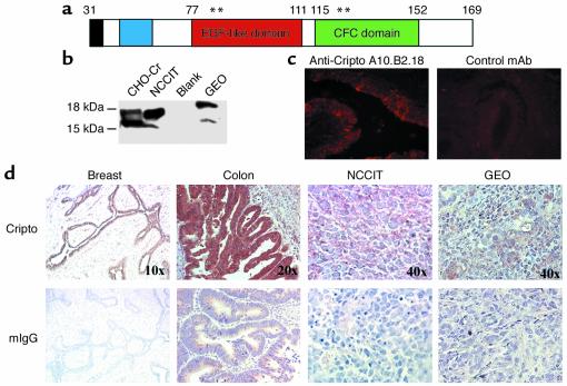
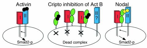
Cripto, a cell surface-associated protein belonging to the EGF-CFC family of growth factor-like molecules, is overexpressed in many human solid tumors, including 70-80% of breast and colon tumors, yet how it promotes cell transformation is unclear. During embryogenesis, Cripto complexes with Alk4 via its unique cysteine-rich CFC domain to facilitate signaling by the TGF-beta ligand Nodal. We report, for the first time to our knowledge, that Cripto can directly bind to another TGF-beta ligand, Activin B, and that Cripto overexpression blocks Activin B growth inhibition of breast cancer cells. This result suggests a novel mechanism for antagonizing Activin signaling that could promote tumorigenesis by deregulating growth homeostasis. We show that an anti-CFC domain antibody, A8.G3.5, both disrupts Cripto-Nodal signaling and reverses Cripto blockade of Activin B-induced growth suppression by blocking Cripto's association with either Alk4 or Activin B. In two xenograft models, testicular and colon cancer, A8.G3.5 inhibited tumor cell growth by up to 70%. Both Nodal and Activin B expression was found in the xenograft tumor, suggesting that either ligand could be promoting tumorigenesis. These data validate that functional blockade of Cripto inhibits tumor growth and highlight antibodies that block Cripto signaling mediated through its CFC domain as an important class of antibodies for further therapeutic development.


Figures

Figure 1
Figure 1
(a) Diagram of mature hCr protein: N-terminal tip (black), N-terminal region (blue), EGF-like domain (red), and CFC domain (green). Amino acid 169 is N-terminal to the GPI-linkage site. Asterisks indicate positions of mutants described in Table 1. (b) Western blot of Cripto expressed in CHO cells (recombinant) or endogenously expressed in NCCIT and GEO tumor cell lines using A10.B2.18. (c) Immunofluorescent staining of cell surface Cripto on human breast tumor tissue with A10.B2.18 (left) or control antibody 1E6 (right). (d) Immunohistochemical staining of tumor sections with anti-Cripto mAb’s A10.B2.18 (breast and NCCIT) and B3.F6.17 (colon and GEO) or mouse IgG as a negative control. Color variations result from the use of different substrates, either Vector NovaRED or DAB substrate kits (Vector Laboratories Inc.).
Figure 2
Figure 2
Anti-CFC mAb’s block Nodal signaling and Cripto-Alk4 interactions. (a) NCCIT cells were transfected with plasmids expressing (n2)7-luciferase and Nodal (column 1) or (n2)7-luciferase, FAST, and Nodal (columns 2–6). A27.F6.1 was added at 0, 1, 3, 10, or 30 μg/ml for 16 hours before luciferase reading (P < 0.001 for each dose). (b) Human 293-Alk4 cells were incubated with Cr-hFc or Cr-hFc prebound with mAb and assayed for binding by FACS using an anti-hFc PE-conjugated secondary mAb. The mean fluorescence intensity of Cr-hFc/mAb bound to 293-Alk4 cells is represented as a percentage of Cr-hFc binding with no mAb. (c) NCCIT cells were assayed for blocking of Cripto-Nodal signaling by A8.G3.5 at 10 μg/ml (P = 0.002) or 30 μg/ml (P = 0.05) as described in a
Figure 3
Figure 3
Overexpression of Cripto blocks Act B growth suppression of breast epithelial cells. (a) T47D or T47D-hCr cells were grown in media alone (white bars) or media plus 25 ng/ml Act A (black bars) or Act B (gray bars). Proliferation was measured by an MTT assay after 8 days. n = 4; the experiment was repeated at least twice. (b) Lysates of T47D and T47D-hCr cells stimulated with 25 ng/ml of Act A (left panel) or Act B (right panel) for 0, 10, 30, and 60 minutes were immunoblotted with anti–phospho-Smad2 polyclonal antibody. B, blank sample of T47D untreated cells. The arrow points to the phospho-Smad2 band. (c) T47D and T47D-hCr cells were stimulated with 0, 5, 25, or 50 ng/ml Act A, Act B, or EGF for 1 hour and then lysed and probed for p44/42 MAPK.
Figure 4
Figure 4
Cripto binds directly to Act B, and binding is blocked by anti-CFC mAb A8.G3.5. (a) Act A or Act B was immunoprecipitated with Cr-hFc prebound to protein A-Sepharose. Immunoprecipitated protein was immunoblotted using anti–Act A (left panel) or anti–Act B (right panel) mAb’s. Act A and Act B (10 ng) were loaded on each gel for comparison. (b) Act B binds to Cr-hFc in a Biacore assay. Act A (left panels) or B (right panels) was flowed over Biacore chips immobilized with Cr-hFc (top panels) or LTβR-hFc (bottom panels) and assayed for binding by surface plasmon resonance using a Biacore 2000 biosensor system (Biacore Inc.). Nonspecific binding to the blank-flow cell was subtracted from each sensorgram to obtain the specific-binding responses. RU, resonance units. (c) Purified Act B or cell supernatant containing Nodal was immunoprecipitated with Cr-hFc or CrEGFmt-hFc as described in a. (d) For antibody blocking, Cr-hFc was preincubated with 0.1–3 μg of B3.F6.17, A27.F6.1, or A8.G3.5 prior to Act B binding.IP, intraperitoneal. WB, Western blot.
Figure 5
Figure 5
Anti–CFC domain mAb A8.G3.5 restores Act B–induced growth inhibition. (a) T47D or T47D-hCr cells grown in media alone (white bars), media with Act B (light gray bars), or media with Act B plus 20 μg/ml A8.G3.5 (black bars) or 10 μg/ml A8.G3.5 (dark gray bars) were assayed for proliferation after 8 days by an MTT assay. n = 4; the experiment was repeated at least twice. (b) T47D or T47D-hCr cells grown in media alone (white bars), media with Act B (light gray bars), or media with Act B plus 20 μg/ml A27.F6.1 (black bars). n = 4; the experiment was repeated at least twice. (c) T47D-hCr cells were transfected with (n2)7-luciferase and FAST, and then treated with Act B with or without A8.G3.5 or A27.F6.1 at 0, 1, 3, 10, or 30 μg/ml for 16 hours before luciferase reading (P < 0.005 for each dose).
Figure 6
Figure 6
Anti–CFC domain mAb blocks NCCIT and GEO tumor growth in vivo. (a) Response of NCCIT xenograft to A27.F6.1: vehicle control (open triangles), cis-platinum (cross-hatches), 1 mg/kg A27.F6.1 (filled squares), 10 mg/kg A27.F6.1 (open circles). (b) Response of NCCIT xenograft to A8.G3.5: vehicle control (open triangles), cis-platinum (cross-hatches), 10 mg/kg A8.G3.5 (closed triangles). (c) Response of NCCIT xenograft to A10.B2.18: vehicle control (open triangles), cis-platinum (cross-hatches), 1 mg/kg A10.B2.18 (filled squares), 3 mg/kg A10.B2.18 (filled circles). (d) Response of GEO xenograft to A8.G3.5: vehicle control (open triangles), cis-platinum (cross-hatches), 1 mg/kg A8.G3.5 (filled squares), 3 mg/kg A8.G3.5 (open circles), 10 mg/kg A8.G3.5 (filled triangles). (e) Response of GEO cell xenograft to B3.F6.17: vehicle control (filled squares), cis-platinum (cross-hatches), 10 mg/kg B3.F6.17 (open circles). (f) RT-PCR expression analysis of mRNA transcripts for Cripto, Alk4, Nodal, and Act B in GEO, NCCIT, or MCF-7 cells. Controls include samples without reverse transcriptase (–RT) and samples in which template was replaced with H2O. (g) RT-PCR expression analysis of mRNA transcripts for murine Act B or human Nodal in NCCIT xenograft tumor sample.
Figure 7
Figure 7
Model of Cripto’s inhibition of Act B signaling through the Smad2 pathway. Left: Activin (black) signaling via Alk4 (red), ActRII (blue), and Smad2 leads to regulated cell proliferation in many tissues and organs. Right: Cripto-dependent Nodal (yellow) signaling through Smad2 leads to differentiation and patterning in the early embryo. Cripto is represented in green. Center: Cripto inhibits Act B signaling via binding to Alk4 and Act B, resulting in deregulated growth and hyperproliferation. Three possible inhibitory complexes are proposed: Cripto/Act B, Cripto/Alk4, and Cripto/Act B/Alk4. Smad2-p, phospho-Smad2.

Comment in

References

    1. Gibbs JB. Anticancer drug targets: growth factors and growth factor signaling. J. Clin. Invest. 2000;105:9–13. - PMC - PubMed
    1. de Bono JS, Rowinsky EK. The ErbB receptor family: a therapeutic target for cancer. Trends Mol. Med. 2002;8(Suppl. 4):S19–S26. - PubMed
    1. George D. Platelet-derived growth factor receptors: a therapeutic target in solid tumors. Semin. Oncol. 2001;28:27–33. - PubMed
    1. Adamson ED, Minchiotti G, Salomon DS. Cripto: a tumor growth factor and more. J. Cell. Physiol. 2002;190:267–278. - PubMed
    1. Ciardiello F, et al. Inhibition of CRIPTO expression and tumorigenicity in human colon cancer cells by antisense RNA and oligodeoxynucleotides. Oncogene. 1994;9:291–298. - PubMed

Publication types

MeSH terms

Substances