. 2003 Aug;112(4):588-97.
doi: 10.1172/JCI18377.

Thyroid hormone action in the absence of thyroid hormone receptor DNA-binding in vivo

Affiliations

Thyroid hormone action in the absence of thyroid hormone receptor DNA-binding in vivo

Nobuyuki Shibusawa et al. J Clin Invest. 2003 Aug.

Abstract

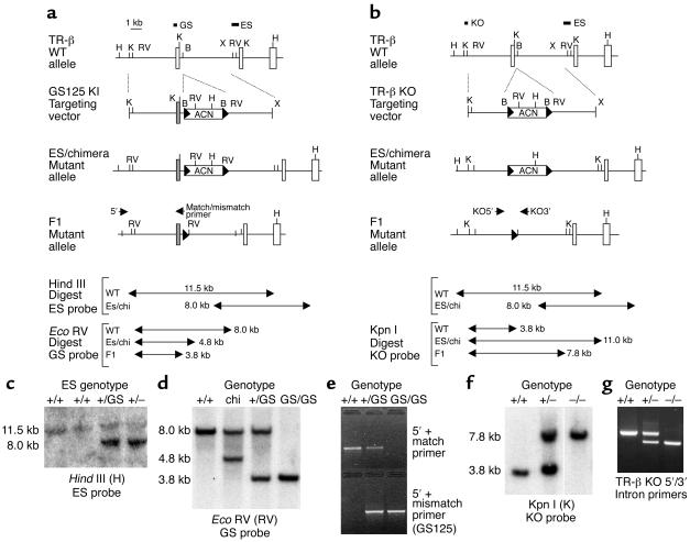
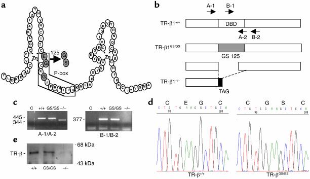
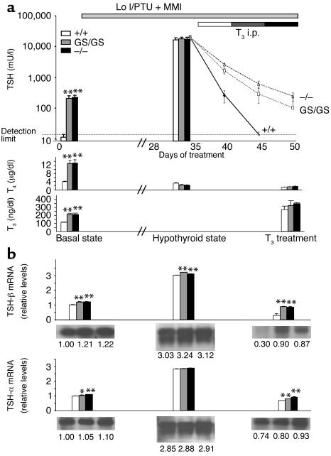
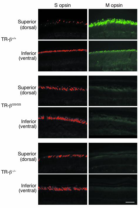
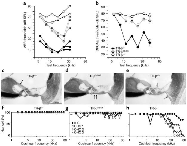
Thyroid hormone action is mediated by thyroid hormone receptors (TRs), which are members of the nuclear hormone receptor superfamily. DNA-binding is presumed to be essential for all nuclear actions of thyroid hormone. To test this hypothesis in vivo, the DNA-binding domain of TR-beta was mutated within its P-box (GS mutant) using gene targeting techniques. This mutation in vitro completely abolishes TR-beta DNA-binding, while preserving ligand (T3) and cofactor interactions with the receptor. Homozygous mutant (TR-betaGS/GS) mice displayed abnormal T3 regulation of the hypothalamic-pituitary-thyroid axis and retina identical to abnormalities previously observed in TR-beta KO (TR-beta-/-) mice. However, TR-betaGS/GS mutant mice maintained normal hearing at certain frequencies and did not display significant outer hair cell loss, in contrast to TR-beta-/- mice. DNA-binding, therefore, is essential for many functions of the TR, including retinal development and negative feedback regulation by thyroid hormone of the hypothalamic-pituitary-thyroid axis. Inner ear development, although not completely normal, can occur in the absence of TR DNA-binding, suggesting that an alternative and perhaps novel thyroid hormone-signaling pathway may mediate these effects.


Figures

Figure 1
Figure 1
Generation of TR-βGS/GS and TR-β–/– mice. Schematic strategy of homologous recombination in TR-βGS/GS (a) and TR-β–/– mice (b) is illustrated. Diagrams show the WT TR-β locus, the targeting vectors, ES-targeted alleles, and the F1 mutant alleles after the ACN cassette is excised. The mutated exon 3 is shown as shaded boxes. H, Hind III; K, Kpn I; RV, Eco RV; B, Bgl II; X, Xba I. The locations of ES, GS, and KO probes for Southern blot analysis are indicated. The ACN cassette is flanked by loxP sites indicated by black arrowheads. Arrows indicate the positions of PCR primers (5′, match/mismatch, KO5′, KO3′) used for DNA genotyping. The site of restriction fragments obtained by Southern blot analysis is given in kb. Chi, chimeric. (c) Southern blot analysis of DNA from ES clones. WT (11.5 kb) and targeted (8.0 kb) Hind III alleles were detected by a 3′ external ES probe. (d) Southern blot analysis of DNA from F2 mice resulting from a GS125 KI heterozygous intercross compared with chimeric animals. After Eco RV digestion, a 4.8-kb band was detected in targeted allele of chimeric mice using GS probe. After self-excision of ACN cassette, a 3.8-kb band is obtained from the mutant allele. (e) Genotyping of F2 KI offspring by mismatch PCR. The WT allele was detected with WT match primer set and mutant allele was detected only with the mismatch (mutant) primer set. (f) Southern blot analysis of TR-β KO F2 siblings using the KO probe. The mutant allele demonstrated a longer (7.8 kb) band after Kpn I digestion versus the WT allele of 3.8 kb. (g) PCR genotyping of F2 KO mice. A 300-bp shorter band is observed in DNA from the KO versus WT allele.
Figure 2
Figure 2
(a) The amino acid sequence of the DNA-binding domain of TR-β. The boxed region outlines the DNA recognition α-helix. The shaded circles indicate the P-box amino acids. The exchange of glutamic acid 125 and glycine 126 to glycine and serine in the GS125 mutation is indicated. (b) Liver RNA was amplified by RT-PCR using primers located in sequences corresponding to exons 2 and 4 (A-1, A-2) and exons 3 and 4 (B-1, B-2), which are indicated by arrows. (c) RT-PCR results from liver RNA using the indicate primers. A 445-bp fragment from WT (+/+) and TR-βGS/GS animals was observed with the A primer set. A short fragment (344 bp) from the A primer set and no band from the B set were obtained from RNA transcripts from TR-β–/– mice. (d) DNA sequence of the 445-bp RT-PCR fragment. The two mutated amino acids are indicated. The deleted mRNA from TR-β–/– encoded a putative peptide that terminated after eight bases in exon 4 at a TAG stop codon. This was confirmed by sequencing the 344-bp fragment from TR-β–/– mice (data not shown). (e) Western blot analysis of liver total cellular protein extracts from WT, TR-β–/–, and TR-βGS/GS animals (a C-terminal TR-β monoclonal antibody was used), indicating that the GS125 mutation did not affect expression from the TR-β locus.
Figure 3
Figure 3
Effect of thyroid hormone deficiency and excess on regulation of the H-P-T axis. (a) Serum TSH levels were sequentially determined in TR-β+/+ (white bar, solid line), TR-βGS/GS (gray bar, dotted line), and TR-β–/– mice (black bar, dashed line) at the baseline, after Lo I/PTU diet+MMI water for 5 weeks, and with the additional treatment with different doses of L-T3 for 5 days each (top). TSH detection limit was less than 25 mU/l. Serum T4 (center) and T3 (bottom) levels were also measured. Five animals were evaluated in each group, and data are shown as means ± SEM. (b) TSH-β (top) and common-α subunit (bottom) mRNA levels in the anterior pituitary of TR-β+/+, TR-βGS/GS, and TR-β–/– mice using a Northern blot analysis. Each Northern blot was rehybridized with cyclophilin probe as a control, and data were normalized for each mRNA level relative to the basal state of WT animals. *P < 0.05, **P < 0.01. i.p., intraperitoneal.
Figure 4
Figure 4
Selective loss of M-cone and altered S-cone distribution in TR-βGS/GS and TR-β–/– mutant mice. The left panels show S opsin immunostaining visualized with red fluorescence. The right panels display M opsin–specific immunostaining labeled with green fluorescence in retina of TR-β+/+, TR-βGS/GS, and TR-β–/– mice. Both superior (dorsal) and inferior (ventral) regions are displayed from each genotype. TR-βGS/GS and TR-β–/– mice showed S opsin expression in significantly more dorsal regions of cone photoreceptors compared with TR-β+/+ mice and had an absence of M opsin expression throughout the retina.
Figure 5
Figure 5
Cochlear function and histopathology in TR-β mutant animals. (a) ABR thresholds for TR-βGS/GS and TR-β–/– mice are elevated with respect to TR-β+/+, but loss is greater in TR-β–/–. Data from individual 8-week-old animals are shown. (b) DPOAE thresholds for TR-βGS/GS and TR-β–/– mice are elevated with respect to WT; however, TR-β–/– exhibited a much greater deficit. Data are mean ± SEM. Group sizes were 10, 18, and 14 for TR-β+/+, TR-βGS/GS, and TR-β–/–, respectively. (ce) Photomicrographs of the upper basal turn (cochlear frequency approximately 16 kHz) from WT and TR-β mutant cochleas. Arrows indicate: (c) tectorial membrane in TR-β+/+; (d) misaligned feet of outer pillar cells in TR-βGS/GS; (e) collapse of outer supporting cells (unfilled) and loss of spiral ligament fibrocytes in TR-β–/– (filled). Scale bar in c applies to all three images. (fh) Basal-turn OHC loss is seen in TR-β–/– mice. Data from one ear of each genotype are shown. Symbol key in g applies to all three panels.

Comment in

Similar articles

Cited by

References

    1. Brent GA. The molecular basis of thyroid hormone action. N. Engl. J. Med. 1994;331:847–853. - PubMed
    1. Oppenheimer JH, Schwartz HL. Molecular basis of thyroid hormone-dependent brain development. Endocr. Rev. 1997;18:462–475. - PubMed
    1. Koibuchi N, Chin WW. Thyroid hormone action and brain development. Trends Endocrinol. Metab. 2000;11:123–128. - PubMed
    1. Cohen, R.C., Weintraub, B.D., and Wondisford, F.E. 2000. Chemistry and biosynthesis of thyrotropin. In Werner and Ingbar’s The thyroid: a fundamental and clinical text. 8th edition. L.E. Braverman, and R.D. Utiger, editors. Williams & Wilkins. Baltimore, Maryland, USA. 202–218.
    1. Forrest D, Erway LC, Ng L, Altschuler R, Curran T. Thyroid hormone receptor beta is essential for development of auditory function. Nat. Genet. 1996;13:354–357. - PubMed

Publication types

MeSH terms