. 2003 Aug 18;162(4):535-41.
doi: 10.1083/jcb.200212107.

Regulation of insulin-like growth factor-dependent myoblast differentiation by Foxo forkhead transcription factors

Affiliations

Regulation of insulin-like growth factor-dependent myoblast differentiation by Foxo forkhead transcription factors

Marta L Hribal et al. J Cell Biol. .

Abstract

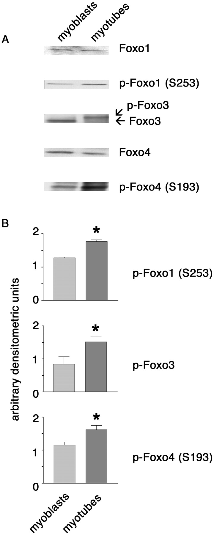
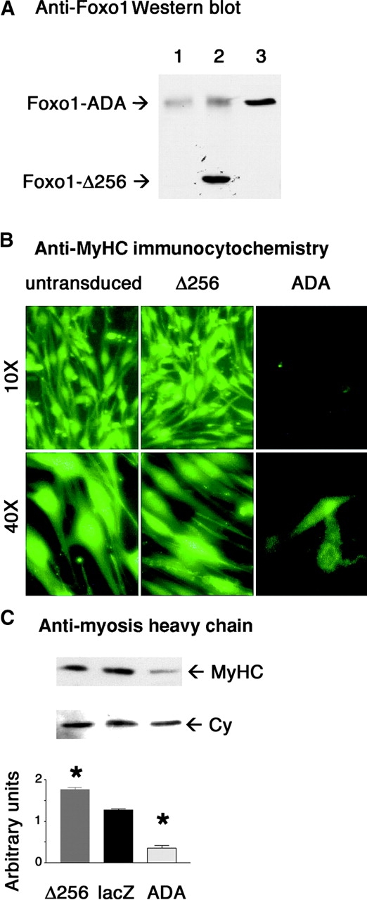
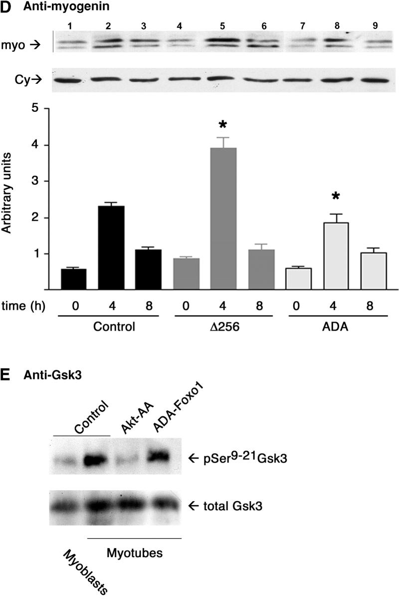
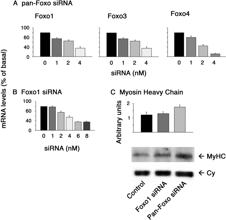
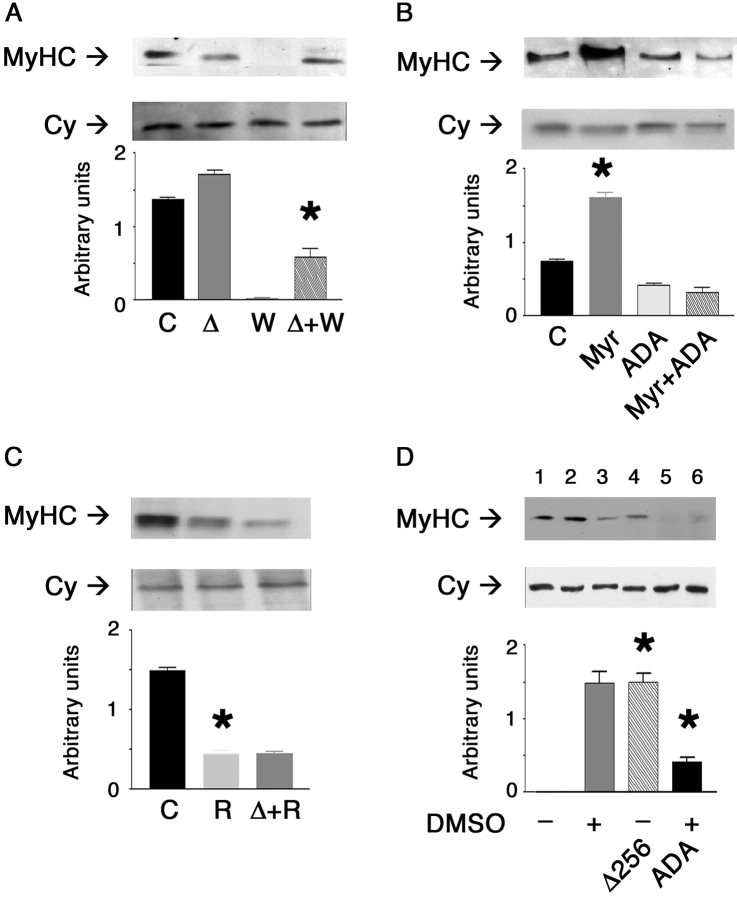
Insulin-like growth factors promote myoblast differentiation through phosphoinositol 3-kinase and Akt signaling. Akt substrates required for myogenic differentiation are unknown. Forkhead transcription factors of the forkhead box gene, group O (Foxo) subfamily are phosphorylated in an insulin-responsive manner by phosphatidylinositol 3-kinase-dependent kinases. Phosphorylation leads to nuclear exclusion and inactivation. We show that a constitutively active Foxo1 mutant inhibits differentiation of C2C12 cells and prevents myotube differentiation induced by constitutively active Akt. In contrast, a transcriptionally inactive mutant Foxo1 partially rescues inhibition of C2C12 differentiation mediated by wortmannin, but not by rapamycin, and is able to induce aggregation-independent myogenic conversion of teratocarcinoma cells. Inhibition of Foxo expression by siRNA resulted in more efficient differentiation, associated with increased myosin expression. These observations indicate that Foxo proteins are key effectors of Akt-dependent myogenesis.


Figures

Figure 1.
Figure 1.
Foxo isoform expression and phosphorylation levels in C 2 C12 myoblasts and myotubes. (A) Extracts of C2C12 myoblasts and myotubes were immunoblotted with the antisera indicated on the right side. (B) Levels of phosphorylated Foxo1 and Foxo4 were assessed by immunoblotting with phospho-specific antisera (S253 for Foxo1, S193 for Foxo4), whereas electrophoretic gel mobility shift was used as a surrogate measure of Foxo3 phosphorylation. The phosphorylated protein migrates more slowly on SDS-PAGE. Mean ± SEM of densitometric scanning values was calculated from three independent experiments. Immunoblotting with anti-cyclophilin antiserum was used as a control for gel loading (not depicted). Asterisk indicates P < 0.01.
Figure 2.
Figure 2.
Expression of early and late differentiation markers in C 2 C12 transduced with Foxo1 mutants. (A) Expression of Foxo1 mutants following adenovirus-mediated gene transfer. Extracts from control cells (lane 1) or from cells transduced with D256-Foxo1 (lane 2) or ADA-Foxo1 (lane 3) were analyzed by immunoblotting with anti-Foxo1 antiserum. (B) Detection of myosin expression by immunocytochemistry. (C) Immunoblot analysis of MyHC expression in cells expressing ADA-Foxo1 or Δ256-Foxo1. A representative blot is shown at the top, and a graph summarizing several experiments is shown on the bottom. The same filter was stripped and reprobed with anti-cyclophilin antiserum (Cy) to normalize protein content (bottom). Asterisk indicates P < 0.01. (D) Myogenin expression in cells expressing mutant Foxo1. Extracts were obtained from untransduced cells (lanes 1–3), cells expressing Δ256-Foxo1 (lanes 4–6), and ADA-Foxo1 (lanes 7–9) at the indicated time points and analyzed by SDS-PAGE, followed by immunoblot with anti-myogenin (top) or anti-cyclophilin antiserum (bottom). Data from three separate experiments are summarized in the bar graphs at the bottom. Scanning densitometry of the autoradiograms was used to measure MyHC and myogenin expression. Data are plotted as mean ± SEM. An asterisk indicates P < 0.01. (E) Phosphorylation of Gsk3 in cells transduced with ADA-Foxo1. Lysates were analyzed by Western blotting with a phosphoSer9–21-specific Gsk3 antiserum (top) and a total Gsk3 antiserum (bottom).
Figure 2.
Figure 2.
Expression of early and late differentiation markers in C 2 C12 transduced with Foxo1 mutants. (A) Expression of Foxo1 mutants following adenovirus-mediated gene transfer. Extracts from control cells (lane 1) or from cells transduced with D256-Foxo1 (lane 2) or ADA-Foxo1 (lane 3) were analyzed by immunoblotting with anti-Foxo1 antiserum. (B) Detection of myosin expression by immunocytochemistry. (C) Immunoblot analysis of MyHC expression in cells expressing ADA-Foxo1 or Δ256-Foxo1. A representative blot is shown at the top, and a graph summarizing several experiments is shown on the bottom. The same filter was stripped and reprobed with anti-cyclophilin antiserum (Cy) to normalize protein content (bottom). Asterisk indicates P < 0.01. (D) Myogenin expression in cells expressing mutant Foxo1. Extracts were obtained from untransduced cells (lanes 1–3), cells expressing Δ256-Foxo1 (lanes 4–6), and ADA-Foxo1 (lanes 7–9) at the indicated time points and analyzed by SDS-PAGE, followed by immunoblot with anti-myogenin (top) or anti-cyclophilin antiserum (bottom). Data from three separate experiments are summarized in the bar graphs at the bottom. Scanning densitometry of the autoradiograms was used to measure MyHC and myogenin expression. Data are plotted as mean ± SEM. An asterisk indicates P < 0.01. (E) Phosphorylation of Gsk3 in cells transduced with ADA-Foxo1. Lysates were analyzed by Western blotting with a phosphoSer9–21-specific Gsk3 antiserum (top) and a total Gsk3 antiserum (bottom).
Figure 3.
Figure 3.
siRNA-mediated inhibition of Foxo expression. Expression of mRNA encoding the three Foxo isoforms in cells transfected with increasing concentrations of pan-Foxo (A) or Foxo1-specific siRNA (B). Results are expressed as a percentage of control. (C) Immunoblot analysis of MyHC expression in cells transfected with pan-Foxo (light gray), Foxo1-specific siRNA (dark gray), or control siRNA (black, top). The same filter was stripped and reprobed with anti-cyclophilin antiserum (Cy) to normalize protein content (bottom).
Figure 4.
Figure 4.
Signaling pathways involved in Foxo1 regulation of myoblast differentiation. (A) Δ256-Foxo1 partly rescues wortmannin inhibition of myoblast differentiation. C, control cells; W, wortmannin-treated cells; Δ, cells expressing Δ256-Foxo1; Δ + W, Δ 256-expressing cells that have been treated with wortmannin. Extracts were obtained 96 h after the induction of differentiation. The bar graph summarizes the results of three experiments. (B) ADA-Foxo1 overrides the effect of Myr-Akt to stimulate myoblast differentiation. Extracts from control cells (C) and cells transduced with Myr-Akt (Myr), ADA-Foxo1 (ADA), or both (Myr + ADA) were prepared 96 h after the induction of differentiation. Equal amounts of protein were subjected to SDS-PAGE and were immunoblotted with anti-MyHC (top) or anti-cyclophilin antiserum (bottom). (C) Δ256-Foxo1 fails to override the inhibitory effect of rapamycin on C2C12 differentiation. Immunoblot analyses with anti-MyHC (top) and anti-cyclophilin (bottom) antisera were performed on extracts obtained from control (C), rapamycin-treated (R), and Δ256-expressing cells (Δ + R) as indicated above. (D) Δ256-Foxo1 induces myogenic conversion of P19 teratocarcinoma cells independently of DMSO treatment. P19 cells were differentiated in the presence (lane 1 and lane 4) or absence (lane 2 and lane 3) of DMSO and were transduced with either Δ256-Foxo1 (lane 2) or ADA-Foxo1 (lane 4). Lane 5 and lane 6 show control RD cells and RD transduced with Δ256-Foxo1. The bar graphs represent mean ± SEM densitometric values from three or four experiments for each condition. One asterisk indicates P < 0.01 in A–C, and P < 0.005 in D. Cyclophilin was used to normalize gel loading.

Similar articles

Cited by

References

    1. Astolfi, A., C. De Giovanni, L. Landuzzi, G. Nicoletti, C. Ricci, S. Croci, L. Scopece, P. Nanni, and P.L. Lollini. 2001. Identification of new genes related to the myogenic differentiation arrest of human rhabdomyosarcoma cells. Gene. 274:139–149. - PubMed
    1. Bois, P.R., and G.C. Grosveld. 2003. FKHR (FOXO1a) is required for myotube fusion of primary mouse myoblasts. EMBO J. 22:1147–1157. - PMC - PubMed
    1. Brunet, A., A. Bonni, M.J. Zigmond, M.Z. Lin, P. Juo, L.S. Hu, M.J. Anderson, K.C. Arden, J. Blenis, and M.E. Greenberg. 1999. Akt promotes cell survival by phosphorylating and inhibiting a forkhead transcription factor. Cell. 96:857–868. - PubMed
    1. Brunet, A., J. Park, H. Tran, L.S. Hu, B.A. Hemmings, and M.E. Greenberg. 2001. Protein kinase SGK mediates survival signals by phosphorylating the forkhead transcription factor FKHRL1 (FOXO3a). Mol. Cell. Biol. 21:952–965. - PMC - PubMed
    1. Canicio, J., E. Gallardo, I. Illa, X. Testar, M. Palacin, A. Zorzano, and P. Kaliman. 1998. p70 S6 kinase activation is not required for insulin-like growth factor-induced differentiation of rat, mouse, or human skeletal muscle cells. Endocrinology. 139:5042–5049. - PubMed

Publication types