. 2003 Aug 18;162(4):565-74.
doi: 10.1083/jcb.200305013.

Visualization of chromatin domains created by the gypsy insulator of Drosophila

Affiliations

Visualization of chromatin domains created by the gypsy insulator of Drosophila

Keith Byrd et al. J Cell Biol. .

Abstract

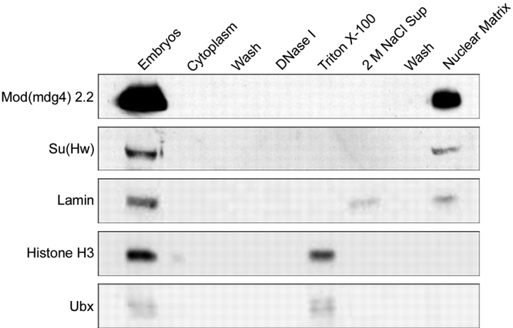
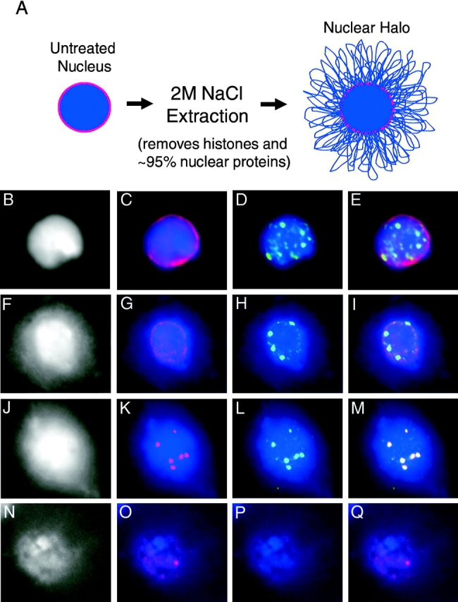
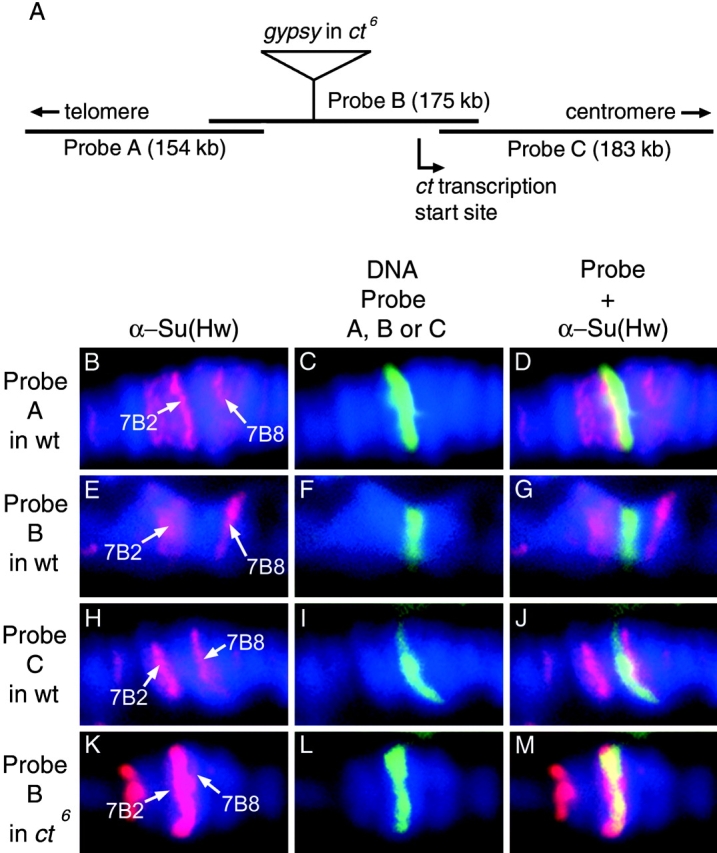
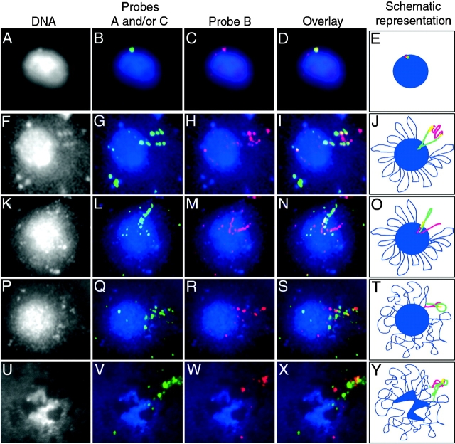
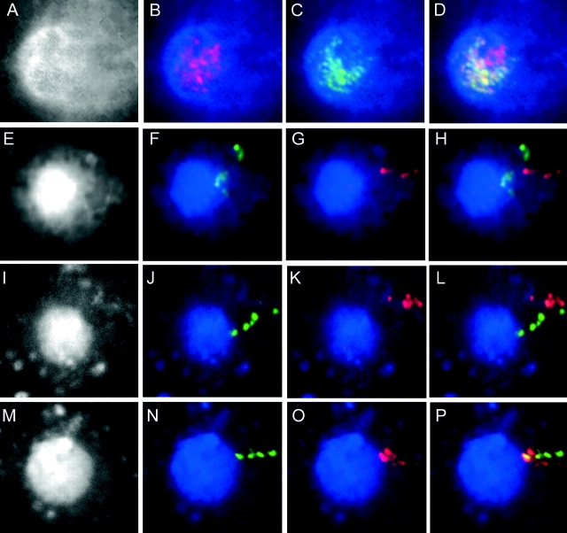
Insulators might regulate gene expression by establishing and maintaining the organization of the chromatin fiber within the nucleus. Biochemical fractionation and in situ high salt extraction of lysed cells show that two known protein components of the gypsy insulator are present in the nuclear matrix. Using FISH with DNA probes located between two endogenous Su(Hw) binding sites, we show that the intervening DNA is arranged in a loop, with the two insulators located at the base. Mutations in insulator proteins, subjecting the cells to a brief heat shock, or destruction of the nuclear matrix lead to disruption of the loop. Insertion of an additional gypsy insulator in the center of the loop results in the formation of paired loops through the attachment of the inserted sequences to the nuclear matrix. These results suggest that the gypsy insulator might establish higher-order domains of chromatin structure and regulate nuclear organization by tethering the DNA to the nuclear matrix and creating chromatin loops.


Figures

Figure 1.
Figure 1.
Protein components of the gypsy insulator are present in the nuclear matrix. A collection of Drosophila 6–18-h-old embryos was subjected to a karyoskeletal or nuclear matrix fractionation procedure, and equal amounts of the fractions were run on 7.5 and 10% polyacrylamide gels and subjected to Western blot analysis using antibodies to Mod(mdg4), Su(Hw), lamin, histone H3, and Ubx. Antibodies against Mod(mdg4) were specific to the Mod(mdg4) 2.2 isoform, which is the isoform present in the gypsy insulator (Ghosh et al., 2001; Mongelard et al., 2002).
Figure 2.
Figure 2.
Distribution of Su(Hw) and Mod(mdg4) insulator proteins after a 2 M NaCl extraction of nuclei. Imaginal disk cells from wild-type larvae were spun onto coverslips and either extracted with 2 M NaCl or left untreated, and then they were stained with antibodies to Su(Hw), Mod(mdg4), or lamin; in addition, the DNA was stained with DAPI (blue). (A) Schematic representation of a nucleus before and after extraction with 2 M NaCl. The red depicts the nuclear lamin and the blue indicates DNA. Treatment of these cells with 2 M NaCl removes histones and extracts ∼95% of nuclear proteins, leaving the DNA in large 50–200-kb loops attached to the residual nuclear matrix (McCready et al., 1979; Ma et al., 1999). B, F, J, and N are black and white images of DNA, corresponding to the panels on their right, visualized by DAPI staining. This staining is shown in blue in the rest of the panels. (C–E) Distribution of lamin (red) and Mod(mdg4)2.2 (green) in untreated nuclei. (G–I) Distribution of lamin (red) and Mod(mdg4)2.2 (green) in 2 M NaCl- extracted nuclei. (K–M) Distribution of Su(Hw) (red) and Mod(mdg4)2.2 (green) in 2 M NaCl-extracted nuclei. (O–Q) Distribution of Su(Hw) (red) and Mod(mdg4)2.2 (green) in nuclei extracted with 2 M NaCl followed by treatment with RNase A.
Figure 3.
Figure 3.
Localization of DNA probes A, B, and C and Su(Hw) protein on polytene chromosomes. BAC clones A, B, or C were used as DNA probes for FISH of polytene chromosomes from salivary glands of Drosophila third instar larvae from wild-type or ct 6 mutant strains. The chromosomes were simultaneously stained with antibodies against the Su(Hw) protein. For all images, the DNA is visualized by DAPI staining (blue), and the locations of endogenous Su(Hw) binding sites at 7B2 and 7B8, indicated by Su(Hw) staining (red), are labeled with arrows. (A) Schematic representation of probes A, B, and C at the ct locus. The ct 6 mutant contains a copy of the gypsy retrotransposon located in the region recognized by probe B. The location of the gypsy retrotransposon and overlap of probes A, B, and C are drawn to scale. (B–D) Immunolocalization of Su(Hw) (red) and FISH signal of probe A in polytene chromosomes from wild-type larvae. (E–G) Immunolocalization of Su(Hw) (red) and FISH signal of probe B (green) in polytene chromosomes from wild-type larvae. (H–J) Immunolocalization of Su(Hw) (red) and FISH signal of probe C (green) in polytene chromosomes from wild-type larvae. (K–M) Immunolocalization of Su(Hw) (red) and FISH signal of probe B in polytene chromosomes from ct 6 larvae.
Figure 4.
Figure 4.
Effect of the gypsy insulator on the distribution of DNA in 2 M NaCl-extracted nuclei from male larvae. Various combinations of DNA probes A, B, and C from chromosomal subdivision 7B (see Fig. 3) were hybridized to imaginal disk cells that were spun onto coverslips and either extracted with 2 M NaCl or left untreated. Probes A and C are in green and probe B is in red. A, F, K, P, and U are black and white images of DNA visualized by DAPI staining. This staining is shown in blue in the rest of the panels. The red depicts probe B, and the green depicts probes A and C in panels G–I and probe A in the rest. E, J, O, T and Y show schematic representations of the results found to their left. (A–D) Probes A (green) and B (red) in an untreated wild-type nucleus. (F–I) Probes A (green), B (red), and C (green) in a 2 M NaCl-extracted wild-type nucleus. (K–N) Probes A (green) and B (red) in a 2 M NaCl-extracted nucleus from larvae carrying the ct 6 mutation. (P–S) Probes A (green) and B (red) in a 2 M NaCl- extracted nucleus from larvae of the genotype ct 6; su(Hw)V. (U–X) Probes A (green) and B (red) in a 2 M NaCl- extracted nucleus also treated with RNase A.
Figure 5.
Figure 5.
Insulator-mediated loop organization after heat shock and in nuclei from female larvae. (A–D) Immunofluorescence analysis of 2 M NaCl- extracted nuclei from imaginal disk cells of ct 6 male larvae subjected to a 30-min heat shock at 37°C using antibodies against Su(Hw) and Mod(mdg4)2.2. The Su(Hw) protein is shown in red and the Mod(mdg4)2.2 protein in green. DNA was stained with DAPI and is shown in A; DAPI staining is shown in blue in B–D. The rest of the panels in the figure show FISH analysis of 2 M NaCl- extracted nuclei from imaginal disk cells using probes A (green) and B (red); in all panels, DAPI staining is shown in gray (E, I, and M) or in blue. (E–H) Nuclei from ct 6 male cells subjected to a 30-min heat shock at 37°C. (I–L) Nuclei from imaginal disk cells of wild-type female larvae. (M–P) Nuclei from imaginal disk cells of ct 6 female larvae.

Similar articles

Cited by

References

    1. Bell, A.C., A.G. West, and G. Felsenfeld. 2001. Insulators and boundaries: versatile regulatory elements in the eukaryotic genome. Science. 291:447–450. - PubMed
    1. Berger, S.L. 2002. Histone modifications in transcriptional regulation. Curr. Opin. Genet. Dev. 12:142–148. - PubMed
    1. Blanton, J., M. Gaszner, and P. Schedl. 2003. Protein:protein interactions and the pairing of boundary elements in vivo. Genes Dev. 17:664–675. - PMC - PubMed
    1. Cai, H.N., and P. Shen. 2001. Effects of cis arrangement of chromatin insulators on ennhancer-blocking activity. Science. 291:493–495. - PubMed
    1. Caron, H., B. van Schaik, M. van der Mee, F. Baas, G. Riggins, P. van Sluis, M.C. Hermus, R. van Asperen, K. Boon, P.A. Voute, et al. 2001. The human transcriptome map: clustering of highly expressed genes in chromosomal domains. Science. 291:1289–1292. - PubMed

Publication types