. 2003 Aug 18;162(4):719-30.
doi: 10.1083/jcb.200211011.

Ankyrin binding mediates L1CAM interactions with static components of the cytoskeleton and inhibits retrograde movement of L1CAM on the cell surface

Affiliations

Ankyrin binding mediates L1CAM interactions with static components of the cytoskeleton and inhibits retrograde movement of L1CAM on the cell surface

Orlando D Gil et al. J Cell Biol. .

Abstract

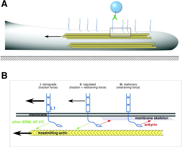
The function of adhesion receptors in both cell adhesion and migration depends critically on interactions with the cytoskeleton. During cell adhesion, cytoskeletal interactions stabilize receptors to strengthen adhesive contacts. In contrast, during cell migration, adhesion proteins are believed to interact with dynamic components of the cytoskeleton, permitting the transmission of traction forces through the receptor to the extracellular environment. The L1 cell adhesion molecule (L1CAM), a member of the Ig superfamily, plays a crucial role in both the migration of neuronal growth cones and the static adhesion between neighboring axons. To understand the basis of L1CAM function in adhesion and migration, we quantified directly the diffusion characteristics of L1CAM on the upper surface of ND-7 neuroblastoma hybrid cells as an indication of receptor-cytoskeleton interactions. We find that cell surface L1CAM engages in diffusion, retrograde movement, and stationary behavior, consistent with interactions between L1CAM and two populations of cytoskeleton proteins. We provide evidence that the cytoskeletal adaptor protein ankyrin mediates stationary behavior while inhibiting the actin-dependent retrograde movement of L1CAM. Moreover, inhibitors of L1CAM-ankyrin interactions promote L1CAM-mediated axon growth. Together, these results suggest that ankyrin binding plays a crucial role in the anti-coordinate regulation of L1CAM-mediated adhesion and migration.


Figures

Figure 1.
Figure 1.
Wild-type or mutant rat L1CAM is expressed on the surface of ND7 cell lines. ND7 cells expressing cDNA constructs encoding either wild-type (B) or mutant L1CAM (C and D), tagged with a myc epitope. Wild-type myc-tagged L1CAM (B) detected by indirect immunofluorescence using an anti-myc antibody (9E10) appears on the cell surface with a distribution that is similar to that of endogenous L1CAM expressed by the cell line (A; rabbit polyclonal against L1CAM). Mutant forms of L1CAM encoding single aa substitutions at tyrosine 1229 including Y1229F (C) and Y1229H (D) are similarly indistinguishable in distribution from endogenous protein. ND-7 cells also express ankyrin B as detected by immunofluorescence (E) and immunoblot (not depicted). (F) Control image of cells expressing wild-type L1CAM stained with secondary antibody alone. Bar, 10 μm.
Figure 2.
Figure 2.
L1CAM–cytoskeleton interactions depend on L1CAM cross-linking. 1-μm latex microspheres coated with anti-myc antibodies recognize cell surface L1CAM tagged with a myc epitope. Bead binding to the cell surface after placement with a laser trap (white bars indicate no binding) depended on antibody concentration. Once bound, beads were tested for resistance to lateral movement with the laser trap (see Materials and methods) as an indication of cytoskeletal attachment. The percentage of trials that are attached and rigid on the cell surface (black bars) or attached and but subject to displacement (gray bars) varied directly with antibody concentration.
Figure 3.
Figure 3.
Wild-type L1CAM engages in three distinct classes of kinetic behavior on the cell surface. 1-μm latex microspheres coated with anti-myc antibodies recognize cell surface L1CAM tagged with a myc epitope. Bound beads underwent diffusion (A, D, and G), retrograde movement (B, E, and H), or stationary behavior (C, F, and I). All data sets were rotated to orient the cell with its leading edge facing left. (A–C) Plots of X vs. Y coordinates of representative data sets (in μm) are consistent with either diffusive (diffusion coefficient D = 0.11 μm2s−1; v = 0 μm min−1), slow directed movement (D = 1.12 × 10⁻⁴ μm2s−1; v = 0.924 μm min−1) or stationary behavior (D = 4.18 × 10⁻⁴ μm2s−1; v = 0 μm s−1; origin indicated by blue arrow). Stationary behavior can be observed as discrete clusters in the bead trajectory (C, black circle). Plots of X and Y coordinates vs. time indicated that the diffusing bead (D) shows no detectable directed movement in the axis of cell migration as movement perpendicular (⊥, fine trace) or parallel (ll, thick trace) to the leading edge are similarly chaotic. In contrast, beads undergoing retrograde movement on the cell surface show slow, uniform movement away from the leading edge with little or no displacement parallel to the leading edge (E). For stationary beads, in the case of the bead shown here (F), diffusive movement decreased (indicated by bars; colors correspond to circles in C after a period of free diffusion [∼10 s]). Mean squared displacement (MSD) for the diffusing bead was linear with respect to time (G), confirming that the behavior is diffusive in the absence of directed movement. The black trace reflects the measured MSD. The red line reflects the best fit of this curve by linear regression. For the retrograde-moving bead (H), the MSD plot has a quadratic shape, reflecting directed movement with substantially reduced diffusion. For stationary beads (I), MSD for the period indicated by the gray bar in F shows no evidence of directed movement.
Figure 4.
Figure 4.
L1CAM–cytoskeleton interactions mediate retrograde and stationary behaviors. Bar graphs showing the percentage of trials engaging in stationary behavior, retrograde movement, or diffusion in cells expressing either wild-type or mutant forms of L1CAM. (A) Wild-type L1CAM treated with actin and microtubule inhibitors. In ND-7 cells expressing wild-type L1CAM, 28.6% of trials were stationary, 61.9% retrograde, and 23.8% diffused (n = 21). Cytochalasin D treatment (2 μM) eliminated retrograde movement (stationary 16.7%, retrograde 0%, diffusing 83.3%; n = 12). In contrast, nocadazole treatment (1 μM) inhibited retrograde movement slightly while leaving stationary behavior and diffusion unaffected (stationary 31.25%, retrograde 43.75%, diffusing 25%; n = 16). Treatment with DMSO alone at the same concentration used to dilute cytochalasin D and nocadazole caused a slight increase in retrograde movement (stationary 22.2%, retrograde 88.8%; diffusing 11.1%; n = 9). (B) L1CAM cytoplasmic tail mutants. In cells expressing an L1 mutant with an introduced stop codon designed to truncate the protein, leaving 4 aa of the predicted cytoplasmic tail (stop), the receptor diffused exclusively. In cells expressing the YF mutant of L1CAM (YF), 62.5% of trials were stationary and 37.5% were retrograde moving (56.2% diffused; n = 16). In cells expressing a histidine in place of Y1229 (FIGQY-H; YH, a mutation that inhibits ankyrin binding), 0% were stationary, 92.9% were retrograde-moving and 7.1% diffusing (n = 14). Finally, in cells expressing wild-type L1CAM treated with NGF to stimulate phosphorylation of Y1229, inhibiting ankyrin binding, 18.2% of trials displayed some form of stationary behavior, whereas 100% underwent retrograde transport on the cell surface (0% diffusing; n = 11).
Figure 5.
Figure 5.
Growth factor treatment inhibits ankyrin B binding to L1CAM. 293 cells transfected with a cDNA encoding full-length rat L1CAM in the absence (A, C, and E) or presence (B, D, and F) of EGF. Ankyrin (A and B) and L1CAM (C and D) were detected by indirect immunofluorescence in double-labeled confocal sections through cell aggregates to permit the visualization of L1CAM and ankyrin B at the cell membrane. (E) Combined micrographs indicating ankyrin staining (red) and L1CAM staining (green) reveal clear codistribution of signal in the absence of EGF. In contrast, in the presence of EGF (F), L1CAM staining remains at the membrane, but ankyrin B staining is largely absent, appearing in a more uniform distribution throughout the cytosol. (G) Direct quantification shows a significant reduction of ankyrin B colocalization with L1CAM at the membrane in the presence of EGF (mean ± SD; P < 0.01). (H) The method for quantifying ankyrin B localization to the membrane uses densitometry of a line scan (red) across a cell–cell junction where L1CAM is expressed (ankyrin B staining is shown here). The resulting intensity profile is used to determine a minimum signal for comparison to the value of the ankyrin B signal at the point where the maximum signal in the L1CAM channel occurs. These values are combined to give an index value using the equation index = (max−min)/min. Index values were averaged and normalized with respect to the control values. Error bars ± SD. Bar, 10 μm.
Figure 6.
Figure 6.
FIGQF peptides inhibit L1CAM–ankyrin interactions, stationary behavior of cell surface L1CAM, and increase the velocity of L1CAM retrograde movement. Peptides derived from the sequence of the ankyrin-binding domain of L1CAM conjugated to the membrane-permeant domain of antennapedia (AP-YF; see text) inhibit L1CAM–ankyrin interactions in membrane recruitment assays (A; P < 0.01). Scrambled peptides (AP-Scramble) where the ankyrin-binding domain is reversed have no detectable effects on L1CAM–ankyrin interactions. (B) Quantification of bead movement shows that the AP-YF peptides inhibit the stationary behavior of beads bound to cell surface L1CAM as compared with treatment with either AP-Scramble peptide or control (untreated) conditions. (C) AP-YF peptides also increase the mean velocity of retrograde movement by approximately twofold as compared with either AP-Scramble (P < 0.01) or control conditions (P < 0.01). A similar increase in the velocity of bead movement is observed using L1CAM mutants that inhibit ankyrin binding (FIGQH; Y1229-H substitution; P < 0.01). Error bars ± SD.
Figure 7.
Figure 7.
Peptide inhibitors of L1CAM–ankyrin interactions selectively stimulate L1CAM-mediated neuronal growth. Cerebellar cells prepared from P4 mouse (1.25 × 10⁵ cells) were plated on chick NgCAM (A and B) or on mouse laminin (C and D). Cultures were fixed after 24 h and images were collected. Cultures were treated with either AP-YF (A and C) or AP-Scramble (B and D) peptides. Measured cell numbers are 105 (laminin + AP-YF), 106 (laminin + AP-Scramble), 322 (NgCAM + AP-YF), and 273 (NgCAM + AP-Scramble). Bar, 125 μm. (E) The percentage of neurons (y-axis, %) with neurites greater than length X (x-axis, μm). Note that treatment of neurons on NgCAM substrates with AP-YF peptide (closed circle) shifted the profile plot to the right compared with control treatment with AP-Scramble peptide (open circle), whereas peptide treatment did not affect profile plot of laminin substrate (squares). Control (BSA) substrate did not promote neurite extension (x). (F) Bar graph showing mean neurite length (μm; error bars represent SEM) for neurons grown under each condition (indicated at bottom).
Figure 8.
Figure 8.
Model describing the regulation of L1CAM dynamics and function by ankyrin binding. (A) Schematic diagram illustrating the leading edge (lamella) of a cell in profile (gray) spread on a two-dimensional surface (black line with crosshatching). Cell is oriented toward the right-hand edge of the page. Treadmilling actin in the lamella of the cell (indicated by yellow chevrons and arrow) provides the force for moving cell surface glycoproteins like L1CAM (light blue) in a retrograde direction on the cell surface. The movement of L1CAM in the plane of the membrane is monitored by beads bound to L1CAM on the upper surface of the cell through a selective antibody (green). (B) Expanded view of area indicated by box in A. Ankyrin-binding domain of L1CAM (light blue) is indicated by oval in cytoplasmic tail. L1CAM–ankyrin interactions mediate the static behavior of L1CAM on the upper surface of the cell. This may be accomplished through ankyrin (red) binding to the membrane/spectrin cytoskeleton. Additionally, L1CAM interacts with dynamic components of the cytoskeleton, presumably treadmilling actin (yellow). These interactions offer a potential explanation for the distinct classes of L1CAM movement that we have observed. The indirect interaction between L1CAM and treadmilling actin in the absence of ankyrin binding results in the retrograde movement of cell surface L1CAM (i). In contrast, ankyrin binding in the absence of interactions with treadmilling actin gives rise to L1CAM that remains stationary on the cell surface (iii). Finally, both interactions together (ii) results in a regulated traction force generation where ankyrin binding modulates the ankyrin-independent generation of traction forces through cell surface L1CAM (smaller black arrow).

Similar articles

Cited by

References

    1. Bretscher, A., K. Edwards, and R.G. Fehon. 2002. ERM proteins and merlin: integrators at the cell cortex. Nat. Rev. Mol. Cell Biol. 3:586–599. - PubMed
    1. Choquet, D., D.P. Felsenfeld, and M.P. Sheetz. 1997. Extracellular matrix rigidity causes strengthening of integrin-cytoskeleton linkages. Cell. 88:39–48. - PubMed
    1. Cohen, N.R., J.S. Taylor, L.B. Scott, R.W. Guillery, P. Soriano, and A.J. Furley. 1998. Errors in corticospinal axon guidance in mice lacking the neural cell adhesion molecule L1. Curr. Biol. 8:26–33. - PubMed
    1. Dahme, M., U. Bartsch, R. Martini, B. Anliker, M. Schachner, and N. Mantei. 1997. Disruption of the mouse L1 gene leads to malformations of the nervous system. Nat. Genet. 17:346–349. - PubMed
    1. Davis, J.Q., and V. Bennett. 1994. Ankyrin binding activity shared by the neurofascin/L1/NrCAM family of nervous system cell adhesion molecules. J. Biol. Chem. 269:27163–27166. - PubMed

Publication types