. 2003 Sep 2;100(18):10417-22.
doi: 10.1073/pnas.1834302100. Epub 2003 Aug 18.

Alzheimer's disease-affected brain: presence of oligomeric A beta ligands (ADDLs) suggests a molecular basis for reversible memory loss

Affiliations

Alzheimer's disease-affected brain: presence of oligomeric A beta ligands (ADDLs) suggests a molecular basis for reversible memory loss

Yuesong Gong et al. Proc Natl Acad Sci U S A. .

Abstract

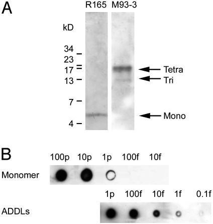
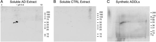
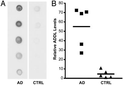
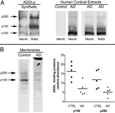
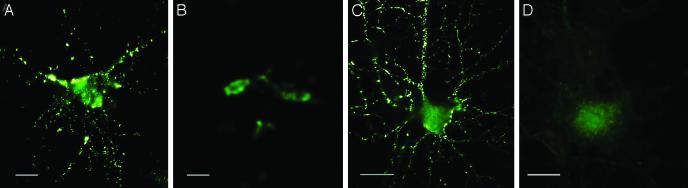
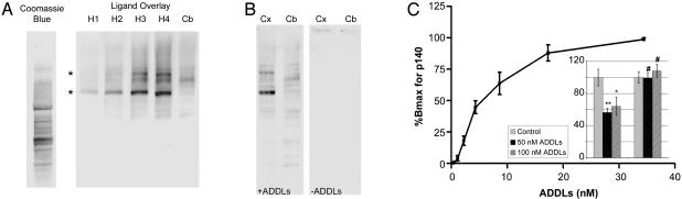
A molecular basis for memory failure in Alzheimer's disease (AD) has been recently hypothesized, in which a significant role is attributed to small, soluble oligomers of amyloid beta-peptide (A beta). A beta oligomeric ligands (also known as ADDLs) are known to be potent inhibitors of hippocampal long-term potentiation, which is a paradigm for synaptic plasticity, and have been linked to synapse loss and reversible memory failure in transgenic mouse AD models. If such oligomers were to build up in human brain, their neurological impact could provide the missing link that accounts for the poor correlation between AD dementia and amyloid plaques. This article, using antibodies raised against synthetic A beta oligomers, verifies the predicted accumulation of soluble oligomers in AD frontal cortex. Oligomers in AD reach levels up to 70-fold over control brains. Brain-derived and synthetic oligomers show structural equivalence with respect to mass, isoelectric point, and recognition by conformation-sensitive antibodies. Both oligomers, moreover, exhibit the same striking patterns of attachment to cultured hippocampal neurons, binding on dendrite surfaces in small clusters with ligand-like specificity. Binding assays using solubilized membranes show oligomers to be high-affinity ligands for a small number of nonabundant proteins. Current results confirm the prediction that soluble oligomeric A beta ligands are intrinsic to AD pathology, and validate their use in new approaches to therapeutic AD drugs and vaccines.


Figures

Fig. 1.
Fig. 1.
Selective, sensitive dot-blot assay for assembled forms of soluble Aβ1-42. (A) Immunoblot of ADDL preparations (100 fmol) shows that M93 antibody selectively identifies oligomers (Right), whereas R165 antibody identifies only monomer (Left). (B) A dot blot immunoassay using M93 detects ADDLs with a sensitivity of 1 fmol, but detects monomers only at levels 1,000-fold higher.
Fig. 3.
Fig. 3.
Assembled forms of soluble Aβ in AD brain show identity with synthetic Aβ oligomers. 2D immunoblots of soluble protein from AD cortex (A) or control cortex (B), and silver stain of synthetic ADDLs (C). AD extract and synthetic ADDLs show a prominent 53-kDa protein at pI 5.6, corresponding to a putative 12-mer of Aβ1-42.
Fig. 2.
Fig. 2.
AD-affected brain is associated with large increases in assembled forms of soluble Aβ.(A) Dot blot immunoassay for assemblies of Aβ in soluble extracts of five AD-affected brains and five age-matched control brains (1 μg of total extracted brain protein per dot). (B) Results from densitometric imaging of these same samples. The line indicates the mean of each set.
Fig. 6.
Fig. 6.
Comparative ligand overlay assays using rat and human membrane proteins. (A Left) Synthetic ligands and rat membranes. Shown is an overlay assay with synthetic Aβ oligomers and rat brain total membranes or fractions enriched in rafts. Additional binding was evident at p100, a band not always detected; compare with Fig. 5A. Binding at p140 and p260 was enriched in rafts. (A Right) Human ligands and rat membranes. Shown is an overlay assay with soluble extracts from human brain (AD or controls) applied to rat brain membranes (total membranes or rafts). AD-soluble extracts showed prominent binding at p100, but binding at p140 and p260 binding was evident in total membranes and in rafts. No signal was found in extracts from control brains. (B Left) synthetic ligands and human membranes. Shown is an overlay assay using total membranes pelleted from human brain and 10 nM synthetic ADDLs. Comparison of control and AD subjects indicated fewer binding sites in AD (note fibrillar amyloid in AD membrane fraction; top of gel). (B Right) Densitometric quantitation of p140 and p260 substantiated fewer binding sites in AD brain.
Fig. 4.
Fig. 4.
ADDLs from AD brain or prepared in vitro show identical punctate binding to neuronal cell-surface proteins. Cultured hippocampal neurons were incubated with soluble extracts of human brain or synthetic ADDLs. Binding was visualized by immunofluorescence microscopy by using M93 antibody. Soluble AD-brain proteins (A), soluble control-brain proteins (B), synthetic ADDLs (C), and synthetic ADDLs (D) pretreated (1 h) with oligomer-selective antibody M71 are shown. Small puncta, typically <1 μm, and largely distributed along neurites, are evident for AD extracts and synthetic ADDLs, but not for control extracts or antibody-pretreated ADDLs. (Bar, 10 μm.)
Fig. 5.
Fig. 5.
ADDLs are high-affinity ligands for particular solubilized proteins from rat brain regions sensitive to ADDL toxicity. (A) Coomassie blue staining or overlay assays using SDS/PAGE-separated membrane proteins from rat hippocampus (H1-4; 12.5, 25, 50, and 75 μg) or cerebellum (Cb; 75 μg). Ligands were synthetic ADDLs (10 nM). Immunodetection used Aβ assembly-dependent rabbit polyclonal antibodies. Selective binding at p140 and p260 kDa bands (*) was readily evident in hippocampus, but not in cerebellum. Binding proteins were not abundant, which was shown by comparison with total hippocampal proteins stained by Coomassie blue. (B) Overlay assays as in A, using 75-μg membrane proteins from rat cortex (Cx) or cerebellum (Cb). Binding at p140 and p260 bands was evident in cortex, but not in cerebellum. Without oligomers (-ADDLs), no signal from the horseradish peroxidase-conjugated secondary antibody was evident. (C) Densitometry of synthetic binding to rat cortical p140, using increasing doses of synthetic ADDLs. Half-maximal binding was at ∼10 nM. (Inset) Impact of ADDLs on MTT reduction in rat cortical (Left) and cerebellar (Right) cultures shows that ADDLs were selectively toxic to cortical cells, with maximal response by 50 nM. Control MTT reduction was taken as 100%.

Similar articles

Cited by

References

    1. Hardy, J. & Selkoe, D. J. (2002) Science 297, 353-356. - PubMed
    1. Kirkitadze, M. D., Bitan, G. & Teplow, D. B. (2002) J. Neurosci. Res. 69, 567-577. - PubMed
    1. Klein, W. L. (2000) in Molecular Mechanisms of Neurodegenerative Diseases, ed. Chesselet, M.-F. (Humana, Totowa, NJ), pp. 1-49.
    1. Kang, J., Lemaire, H. G., Unterbeck, A., Salbaum, J. M., Masters, C. L., Grzeschik, K. H., Multhaup, G., Beyreuther, K. & Muller-Hill, B. (1987) Nature 325, 733-736. - PubMed
    1. Goate, A., Chartier-Harlin, M. C., Mullan, M., Brown, J., Crawford, F., Fidani, L., Giuffra, L., Haynes, A., Irving, N. & James, L. (1991) Nature 349, 704-706. - PubMed

Publication types

MeSH terms