. 2003 Sep 2;100(18):10193-200.
doi: 10.1073/pnas.1733909100. Epub 2003 Aug 18.

A noncanonical sequence phosphorylated by casein kinase 1 in beta-catenin may play a role in casein kinase 1 targeting of important signaling proteins

Affiliations

A noncanonical sequence phosphorylated by casein kinase 1 in beta-catenin may play a role in casein kinase 1 targeting of important signaling proteins

Oriano Marin et al. Proc Natl Acad Sci U S A. .

Abstract

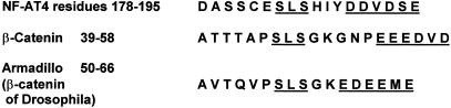
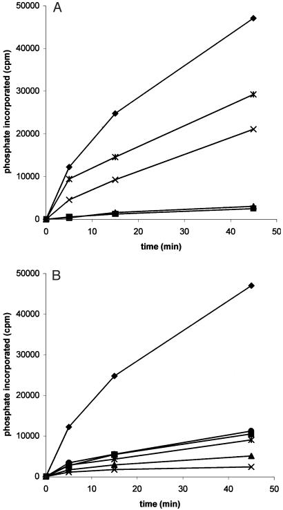
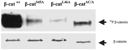
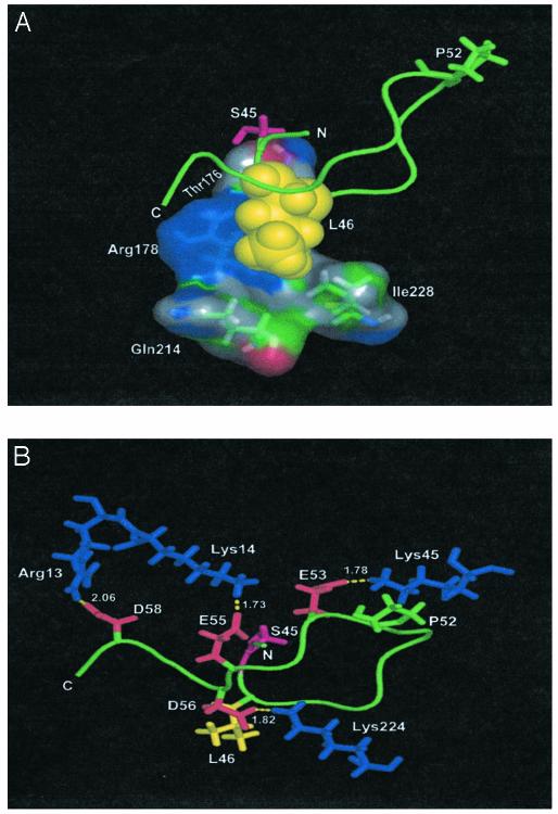
Protein kinase casein kinase 1 (CK1) phosphorylates Ser-45 of beta-catenin, "priming" the subsequent phosphorylation by glycogen synthase-3 of residues 41, 37, and 33. This concerted phosphorylation of beta-catenin signals its degradation and prevents its function in triggering cell division. The sequence around Ser-45 does not conform to the canonical consensus for CK1 substrates, which prescribes either phosphoamino acids or acidic residues in position n-3 from the target serine. However, the beta-catenin sequence downstream from Ser-45 is very similar to a sequence recognized by CK1 in nuclear factor for activated T cells 4. The common features include an SLS motif followed two to five residues downstream by a cluster of acidic residues. Synthetic peptides reproducing residues 38-65 of beta-catenin were assayed with purified rat liver CK1 or recombinant CK1 alpha and CK1 alpha L from zebrafish. The results demonstrate that SLS and acidic cluster motifs are crucial for CK1 recognition. Pro-44 and Pro-52 are also important for efficient phosphorylation. Similar results were obtained with the different isoforms of CK1. Phosphorylation of mutants of full-length recombinant beta-catenin from zebrafish confirmed the importance of the SLS and acidic cluster motifs. A search for proteins with similar motifs yielded, among other proteins, adenomatous polyposis coli, previously found to be phosphorylated by CK1. There is a strong correlation of beta-catenin mutations found in thyroid tumors with the motifs recognized by CK1 in this protein.


Figures

Fig. 1.
Fig. 1.
Alignment of sequences from NF-AT4 (human), β-catenin (human), and Armadillo (the β-catenin homologue of Drosophila) to highlight the similarities that may be recognized by CK1.
Fig. 2.
Fig. 2.
Time course of phosphorylations of β-catenin-derived peptides by CK1. The phosphorylations were carried out as described in Materials and Methods using peptides at 200μM concentration and rat liver CK1. (A) The incidence of the SLS motif is analyzed, with peptide ♦, parent or WT; *, L46V; X, S47A; ▴, L46A, and ▪, S45A. (B) The relevance of the acidic cluster is analyzed, with peptides ♦, parent or WT; *, E53A, E55A, and D58A; X, C terminal (Δ38-48); ▴, N terminal (Δ 52-65); •, E55K; and ▪, acidic all A, a peptide in which E53, D54, E55, D56, and D58 all have been changed to A.
Fig. 3.
Fig. 3.
The phosphorylation of full-length β-catenin WT and mutants by CK1α. The recombinant, bacterially expressed, (His)6-tagged β-catenin WT and mutants from zebrafish were phosphorylated in vitro with the homologous CK1αL enzyme as described in Materials and Methods. The mutants included Ser-45 replaced by alanine (S45A), Leu-46 replaced by alanine (L46A), and all components of the acidic cluster (E53, D54, E55, D56, and D58) changed to alanine (AC/A). (Upper) Bands are autoradiography of the 32P incorporated into β-catenin after SDS/PAGE fractionation. (Lower) Band represents the Western blot of the (His)6 β-catenin present that was developed with monoclonal anti-(His)6 antibody.
Fig. 4.
Fig. 4.
Modelization of the mode of binding of a peptide encompassing the 44-58 sequence of β-catenin to the active site of CK1δ. The backbone of the peptide and the side chain of Pro-52 are shown in green. Phosphorylatable Ser-45 is indicated in magenta. (A) The spheres representing Leu-46 (yellow) are embedded in a cavity formed by four residues of CK1 shown as molecular surface. The minimum distances between peptide Leu-46 and CK1 Thr-176, Arg-178, Gln-214, and Ile-228 are 2.55, 2.64, 3.79, and 3.78 Å, respectively. (B) The interactions among four acidic residues of the peptide (red) clustered in its C-terminal part and four basic residues of CK1 (blue) are highlighted. N and C denote the amino- and carboxyl-terminal ends of the peptide, respectively. The residues of the β-catenin peptide and CK1 are indicated by the one- and three-letter codes, respectively. The β-catenin peptide structure was first built from the amino acid sequence (PS45LSGKGNPEDEDVD) using the protein program of the tinker molecular modeling software (53) with Amber98 force field. The structure was minimized with the program optimize until the energy reached a minimum and manually superimposed to the CK1δ structure (Protein Data Bank ID code 1CK-J.pdb). An energy minimization of the complex CK1-peptide was performed with the program minimize and Amber98 force field until the energy reached a minimum. The figure was drawn by using the pymol program (K. L. DeLano, www.pymol.org).

References

    1. Appella, E. & Anderson, C. W. (2001) Eur. J. Biochem. 268, 2764-2772. - PubMed
    1. Flotow, H., Graves, P. R., Wang, A., Fiol, C. J., Roeske, R. W. & Roach, P. J. (1990) J. Biol. Chem. 265, 14264-14269. - PubMed
    1. Meggio, F., Perich, J. W., Reynolds, E. C. & Pinna, L. A. (1991) FEBS Lett. 283, 303-306. - PubMed
    1. Harwood, A. J. (2001) Cell 105, 821-824. - PubMed
    1. Dajani, R., Fraser, E., Roe, S. M., Young, N., Good, V., Dale, T. C. & Pearl, L. H. (2001) Cell 105, 721-732. - PubMed

Publication types

MeSH terms