. 2003 Aug;14(8):3065-81.
doi: 10.1091/mbc.e02-08-0537. Epub 2003 Jun 13.

Activity and distribution of paxillin, focal adhesion kinase, and cadherin indicate cooperative roles during zebrafish morphogenesis

Affiliations

Activity and distribution of paxillin, focal adhesion kinase, and cadherin indicate cooperative roles during zebrafish morphogenesis

Bryan D Crawford et al. Mol Biol Cell. 2003 Aug.

Abstract

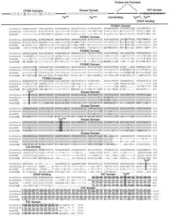
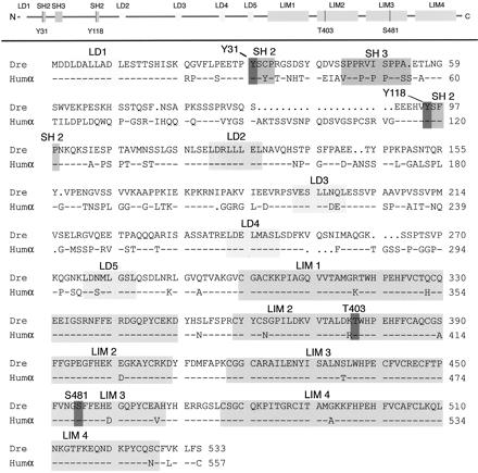
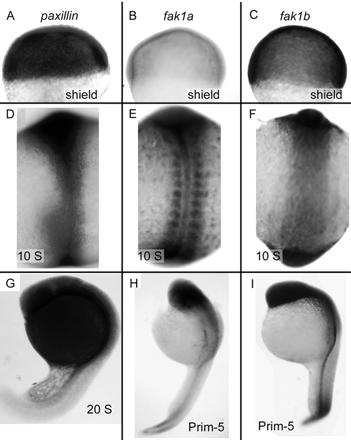
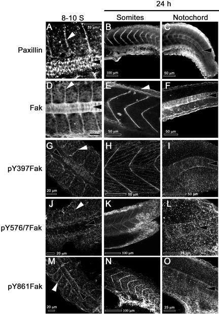
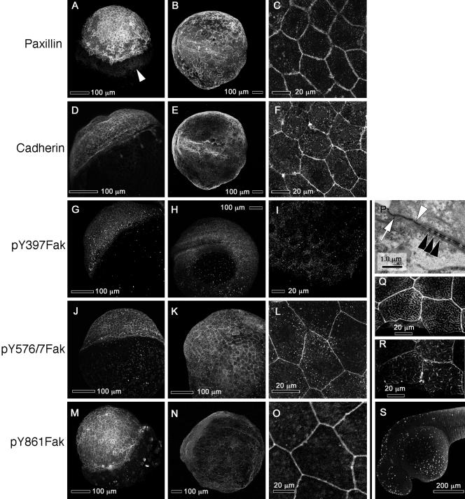
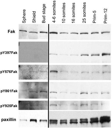
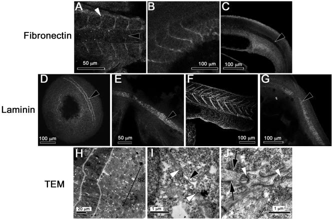
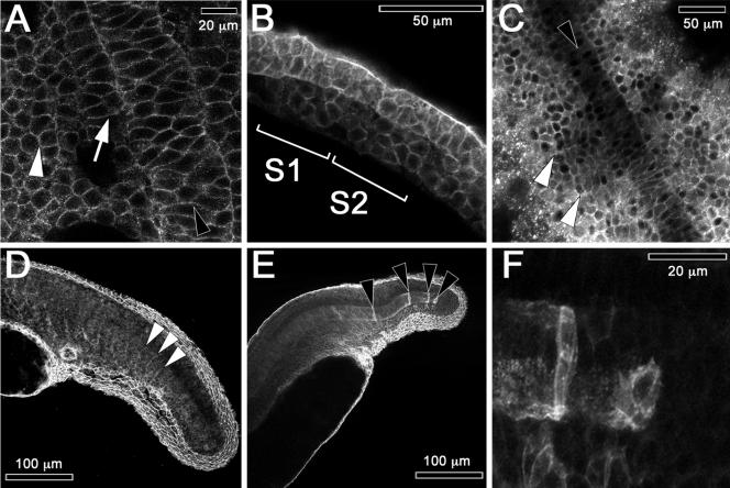
We investigated the focal adhesion proteins paxillin and Fak, and the cell-cell adhesion protein cadherin in developing zebrafish (Danio rerio) embryos. Cadherins are expressed in presomitic mesoderm where they delineate cells. The initiation of somite formation coincides with an increase in the phosphorylation of Fak, and the accumulation of Fak, phosphorylated Fak, paxillin, and fibronectin at nascent somite boundaries. In the notochord, cadherins are expressed on cells during intercalation, and phosphorylated Fak accumulates in circumferential rings where the notochord cells contact laminin in the perichordal sheath. Subsequently, changes in the orientations of collagen fibers in the sheath suggest that Fak-mediated adhesion allows longitudinal expansion of the notochord, but not lateral expansion, resulting in notochord elongation. Novel observations showed that focal adhesion kinase and paxillin concentrate at sites of cell-cell adhesion in the epithelial enveloping layer and may associate with actin cytoskeleton at epithelial junctions containing cadherins. Fak is phosphorylated at these epithelial junctions but is not phosphorylated on Tyr397, implicating a noncanonical mechanism of regulation. These data suggest that Fak and paxillin may function in the integration of cadherin-based and integrin-based cell adhesion during the morphogenesis of the early zebrafish embryo.


Figures

Figure 2.
Figure 2.
Zebrafish Fak1b is conserved with respect to zebrafish Fak1a and human FAK1 and differs significantly from human CAKβ. The FERM, kinase, and the FAT domains are shaded and labeled. The SH2 binding domains have their tyrosine residues darkening. The proline-rich Cas- and GRAF-binding domains are labeled. Dre designates the zebrafish D. rerio sequences and Hum the human sequences. Dre Fak1a is in Henry et al. (2001) and GenBank AAK31154; Dre Fak1b in GenBank AY196213; Hum FAK1 from GenBank Q05397; and Hum CAKβ from GenBank Q14289.
Figure 1.
Figure 1.
Zebrafish paxillin is conserved. The LD and LIM domains, and the SH2 and SH3 binding sites are shaded and labeled. Typically, phosphorylated amino acid residues are shaded and marked as Y31, Y118, T403, and S481. Dre is the zebrafish D. rerio sequence and Humα the α-paxillin isoform of human. D. rerio protein and cDNA sequences are in GenBank AY144487.
Figure 3.
Figure 3.
Expression of paxillin, fak1a, and fak1b mRNAs in the zebrafish embryo. (A–C) Shield stage embryos (6 hpf) hybridized with paxillin (A), fak1a (B), and fak1b (C) probes. Animal pole is up. (D–F) Ten-somite embryos (14 hpf) hybridized with paxillin (D), fak1a (E), and fak1b (F) probes. Presumptive head is up. (G) A 20-somite embryo (19 hpf) hybridized with a paxillin probe (G). (H and I) Prim-5 embryos (24 hpf) hybridized with fak1a (H) and fak1b (I) probes. Head is up and pointing left.
Figure 5.
Figure 5.
Paxillin and Fak are concentrated at somite boundaries and in the notochord, where Fak is phosphorylated at sites of cell-matrix adhesion. The left column of panels shows confocal images of dorsal views of 8- to 10-somite embryos (13.5 hpf, anterior to the left). The middle column shows lateral views of prim-5 embryos (24 hpf, dorsal up) that are focused on the somites. The right column shows lateral views of prim-5 embryos (dorsal up) that are focused on the notochord. The specificities of the antibodies used are indicated at the left. White arrowheads denote somite boundaries and black arrowheads the notochord, except in 5E, where the white arrowhead indicates faint Fak immunoreactivity in the EVL.
Figure 9.
Figure 9.
Paxillin, cadherin, and noncanonically phosphorylated Fak concentrate at the margins of the EVL cells. (A–O) Each row shows confocal images of immunostaining with an antibody against the indicated target. The first column (A, D, G, J, and M) shows embryos at 50% epiboly (5 hpf). The second column (B, E, H, K, and N) shows embryos at the six-somite stage (13 hpf), and the third column (C, F, I, L, and O) shows high-magnification confocal images of the EVL in prim-5 embryos (24 hpf). (A) White arrowhead shows paxillin on the surface of the equatorial region of the yolk. (B and E) Single embryo double-labeled for paxillin and cadherin. (C and F) High-magnification of a single embryo double-labeled for paxillin and cadherin. (P) TEM of a section through the EVL of a prim-5 embryo showing apical tight junctions (white arrow) and adherens junctions (white arrowhead) and desmosomes (black arrowheads) between lateral membranes of EVL cells. (Q and R) High magnifications of confocal images the EVL of a single prim-5 stage embryo showing phalloidin labeling of actin in Q and paxillin labeling in R. The images are near the apical surface of the cell. (S) TUNEL labeling of EVL nuclei in a prim-15 embryo (30 hpf), showing apoptosis. (A and M) Animal poles are up and tilted toward the viewer. (D, G, and J) Animal poles are toward the upper left and slightly away from the viewer. (B, E, and H) Anterior is to the left and dorsal toward the viewer. (K and N) Dorsal is up. (S) Anterior is to the left and dorsal up. (C, F, I, L, and O) Single confocal sections of the EVL. All other panels are confocal projections of Z-stacks.
Figure 4.
Figure 4.
Phosphorylation of Fak and abundance of both paxillin and Fak increase at the end of gastrulation. Each lane represents Western blots from extracts of ten embryos probed with either a polyclonal antibody to the C termini of Fak, or affinity-purified polyclonal antibodies to the phosphorylated SH2 binding sites of Fak, or a monoclonal antibody to paxillin as described in MATERIALS AND METHODS. To maximize detection of antigens before the onset of somitogenesis, the exposures were 7 to 10 times that used for the later stages.
Figure 6.
Figure 6.
Fibronectin is concentrated at somite borders, whereas laminin is concentrated in the forming notochord sheath and at somite borders only after the eight-somite stage. The antigens are given to the left of each row of confocal images. (A) Eight-somite embryo (13 hpf). (B) Prim-5 embryo (24 hpf). (C) Tail of a prim-5 embryo focused on the notochord. (D) Bud-stage embryo (10 hpf), showing staining surrounding the notochord. (E) Eight-somite embryo showing the notochord. (F) Tail of a prim-5 embryo focused on the somites. (G) Prim-5 embryo focused on the notochord. (A, D, and E) Dorsal view, anterior is to the left or left and up. (B, C, F, and G) Lateral view, anterior is to the left. (A–G) Black arrowheads denote the notochord and white arrowheads somite boundaries. (H) Low-magnification TEM of a horizontal section through a three-somite embryo (11 hpf). Two somites are marked (S2 and S3). The notochord is outlined in gray. (I) High-magnification TEM of the section in H focused on a membrane gap with small “island” of matrix forming at the presumptive S2/S3 somite boundary (black arrowhead). Membranes of abutting somite border cells in close contact are marked with white arrowheads. (J) High-magnification TEM of a horizontal section through the somite boundary of a prim-5 embryo (24 hpf) showing rich ECM between somites or black arrows and cellular projections that cross this ECM and form junctions with cells in adjacent somites (white arrowheads).
Figure 7.
Figure 7.
Cadherin concentrates at plasma membranes in somite and notochord cells. Embryos are labeled with anti-cadherin antibody in these confocal images. (A) Bud-stage embryo (10 hpf, dorsal view, anterior up). Intercalating notochord (black arrowhead), adaxial cells (white arrow), and presomitic mesoderm (white arrowhead). (B) Lateral view of a two-somite embryo (10.6 hpf, anterior to the left, dorsal up). Somites 1 and 2 are labeled as S1 and S2, respectively. (C) Eight-somite embryo (13 hpf, dorsal view, anterior up). White arrowheads denote somite boundaries and a black arrowhead the notochord. (D) Lateral view of the tail of a prim-5 embryo (24 hpf) that is focused on formed somites to the left. White arrowheads mark outlines of recently formed somites. (E) A lateral view of the tail of a prim-5 embryo focused on the notochord and the fin-fold. Black arrowheads mark isolated caudal notochord cells expressing high levels of cadherin. (F) High-magnification view of the embryo in E showing the caudal tip of the notochord.
Figure 8.
Figure 8.
Ultrastructure of the notochord sheath reveals patterns of stress that correlate with rings of phosphorylated Fak. (A) TEM of a horizontal section through the notochord of a prim-5 embryo (24 hpf). Black arrowheads indicate changes in the orientation of fibers in the inner layer. The notochord is to the left of the sheath and somites to the right. (B) High-magnification TEM of this notochord sheath showing sites where the membrane of a notochord cell adheres to the fibrous sheath (black arrowheads). (D) Projection of confocal sections of the notochord of a prim-5 embryo stained with anti-pY861Fak shows circumferential striations (anterior up, dorsal to the right). (C) TEM of a horizontal section through the notochord of a three-somite embryo (11 hpf) showing the immature notochord sheath marked with black arrowheads. The notochord cells are to the left of the sheath. (E) High-magnification TEMs of the notochord sheath (black arrowheads) in a three-somite embryo showing only circumferential fiber orientations.

Similar articles

Cited by

References

    1. Adams, D.S., Keller, R., and Ma, K. (1990). The mechanics of notochord elongation, straightening and stiffening in the embryo of Xenopus laevis. Development 110, 115–130. - PubMed
    1. Almeida, E.A.C., Ilic, D., Han, Q., Hauck, C.R., Jin, F., Kawakatsu, H., Schlaepfer, D.D., and Damsky, C.H. (2000). Matrix survival signaling: from fibronectin via focal adhesion kinase to c-Jun NH2-terminal kinase. J. Cell Biol. 149, 741–754. - PMC - PubMed
    1. Avizienyte, E., Wyke, A.W., Jones, R.J., McLean, G.W., Westhoff, M.A., Brunton, V.G., and Frame, M.C. (2002). Src-induced de-regulation of E-cadherin in colon cancer cells requires integrin signaling. Nat. Cell Biol. 4, 632–638. - PubMed
    1. Babb, S.G., Barnett, J., Doedens, A.L., Cobb, N., Liu, Q., Sorkin, B.C., Yelick, P.C., Raymond, P.A., and Marrs, J.A. (2001). Zebrafish E-cadherin: expression during early embryogenesis and regulation during brain development. Dev. Dyn. 22, 231–237. - PubMed
    1. Barrantes, I.B., Elia, A.J., Wunsch, K., De Angelis, M.H., Mak, T.W., Rossant, J., Conlon, R.A., Gossler, A., and de la Pompa, J.L. (1999). Interaction between notch signaling and lunatic fringe during somite boundary formation in the mouse. Curr. Biol. 9, 470–480. - PubMed

Publication types

MeSH terms