. 2003 Aug;14(8):3082-96.
doi: 10.1091/mbc.e02-10-0631. Epub 2003 May 18.

The tumor suppressor cybL, a component of the respiratory chain, mediates apoptosis induction

Affiliations

The tumor suppressor cybL, a component of the respiratory chain, mediates apoptosis induction

Timur Albayrak et al. Mol Biol Cell. 2003 Aug.

Abstract

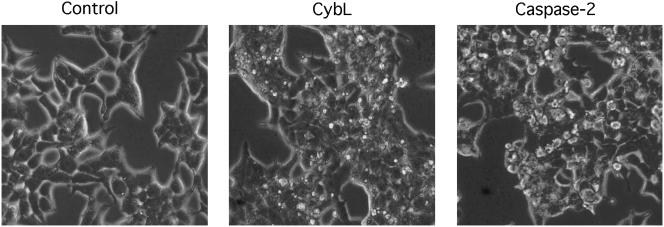
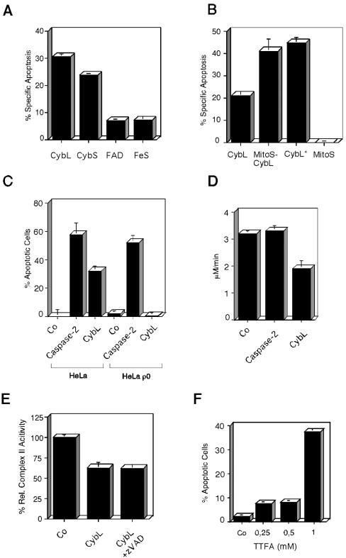
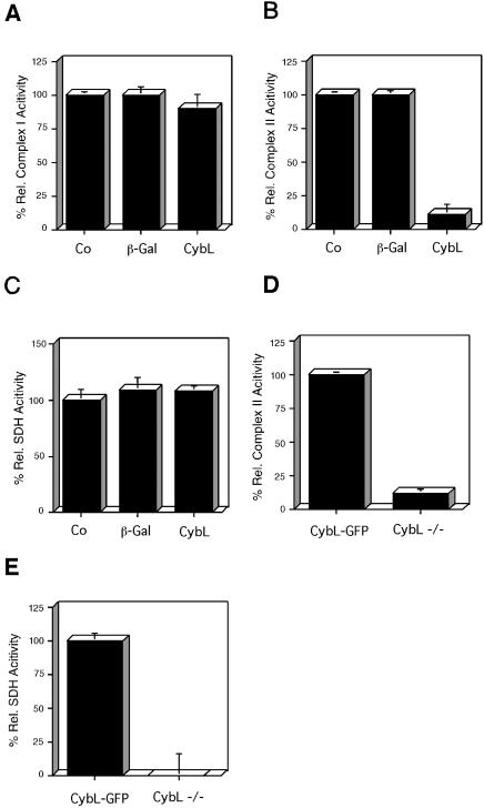
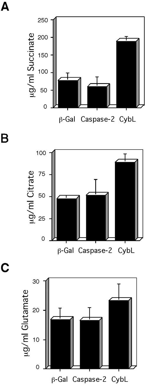
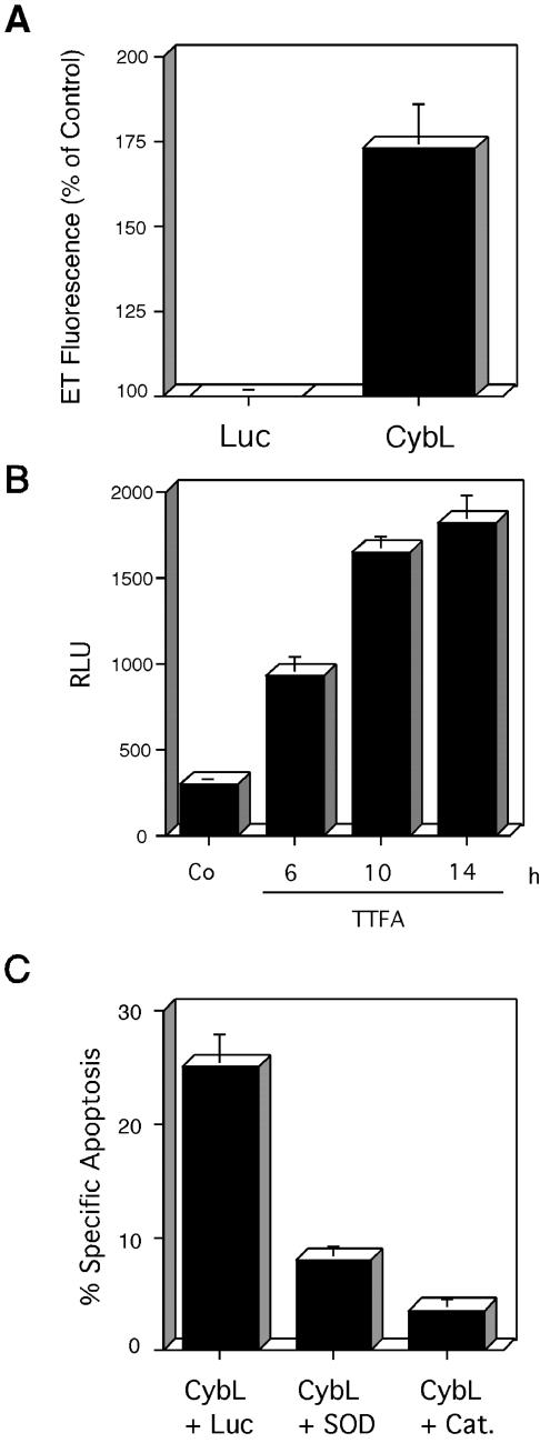
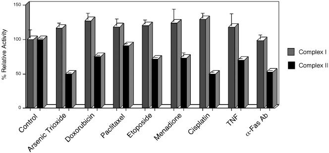
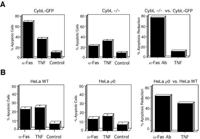
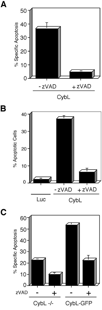
A genetic screen was established to clone apoptosis-inducing genes in a high-throughput format. It led to the isolation of several proapoptotic genes whose proteins are localized to mitochondria. One of the isolated genes is cytochrome bL (cybL also known as SDHC, CII-3, or QPs-1), a component of the respiratory chain complex II. It was further investigated because both cybL and another component of complex II, cybS, have recently been identified as tumor suppressor proteins, some of which act by controlling apoptosis. Our studies reveal that cell death induction by cybL expression is concomitant with a transient inhibition of complex II and the generation of reactive oxygen species. Importantly, cells that are constitutively deficient in cybL are resistant to a variety of proapoptotic cytostatic drugs and to the effects of the Fas receptor. Our results therefore identify complex II as a sensor for apoptosis induction and could explain the unexpected observation that complex II is inactivated in tumors.


Figures

Figure 1.
Figure 1.
Morphological differences between apoptosis induction by caspase-2 and several genes that were isolated with the screen as exemplified by cybL. Expression vectors for cybL and for caspase-2 (2 μg each) were transfected into 293T cells. Phase contrast pictures were taken after 16 h at a 320-fold magnification.
Figure 2.
Figure 2.
Characterization of the proapoptotic stimulus exerted by cybL expression. (A) Test of all components of complex II of the respiratory chain for apoptosis induction. Expression plasmids of cybL and cybS as well as the FAD and the FeS were transfected into 293T cells. Apoptosis was quantified 26 h later by using FACS analysis. (B) Importance of mitochondria for cell death induced by cybL. The isolated cybL isoform was fused at the N terminus to the coding sequence of the mitochondrial import signal of cyclophilin D. Plasmids coding for this fusion protein (MitoS-CybL), the import sequence alone (MitoS), the isolated cybL (CybL) and a longer splice-isoform of cybL (CybL*) were transfected into 293T cells together with a GFP-expression construct. Apoptosis was measured 26 h after starting the transfection by using FACS. Shown are the means and the standard deviations of apoptosis induction of three independent experiments with each construct. (C) Role of the mitochondrial respiratory chain for apoptosis induction by cybL. Wild-type HeLa cells and HeLa ρ0 cells, which harbor mitochondria without mitochondrial DNA and therefore have a defective respiratory chain, were transfected with a control plasmid, an expression vector for cybL or caspase-2, and a GFP construct. Twenty-two hours after starting the transfection the extent of apoptosis in the GFP-positive cells was determined by counting morphologically apoptotic cells. The mean and the SD of three independent results are given for each construct. (D) Effect of cybL expression on the activity of complex II of the respiratory chain. Ten micrograms of an expression plasmid for cybL, for caspase-2, or a control vector was transfected into 10-cm dishes with 293T cells. Thirteen hours after starting the transfection, the mitochondria were isolated and complex II activity of the respiratory chain was assessed with succinate, which reduces dichlorophenylindophenol whose concentration was measured photometrically. For each transfection, a data point was taken every 10 s. Shown are the activities in total cell extracts as the means and SD of three independent experiments for each construct. (E) Effect of cybL expression on complex II activity as measured by an assay with cytochrome c as electron acceptor. Eighteen hours after the transfection of cybL into 293T cells (with and without caspase inhibitor zVAD), mitochondrial extracts were prepared and tested for complex II activity. These measurements were normalized to the transfection efficiency as determined by cotransfected GFP expression vectors. (F) Apoptosis induction by the complex II inhibitor TTFA in HeLa cells. Increasing concentrations of TTFA were added to HeLa cells and apoptosis was detected by FACS analysis.
Figure 3.
Figure 3.
Effect of cybL expression on the activity of different complexes of the respiratory chain. (A–C) The same extracts of untreated 293T cells (Co) or cells transfected with the indicated expression plasmids for β-gal or cybL were investigated for their complex I activity (A), complex II function (B), and SDH (C). Shown are the activities normalized to the transfection efficiencies and relative to the untreated control as means and SD of three independent experiments. (D and E) Complex II activity and succinate dehydrogenase in cybL-negative and -positive cells. (D) Complex II activity was assessed in cybL-negative (cybL –/–) and -reconstituted (cybL-GFP) cells. (E) Succinate dehydrogenase activity was investigated in cybL-negative and -positive cells. Shown are the activities relative to the cybL-reconstituted cells as means and SD of three independent experiments.
Figure 4.
Figure 4.
Consequences of cybL expression on metabolites. The concentrations of succinate (A), citrate (B), and glutamate (C) in the same extracts of 293T cells were determined after transfection of expression plasmids for β-gal, caspase-2, or cybL. Shown are the concentrations normalized to the transfection efficiencies as the means and SD of three independent experiments.
Figure 5.
Figure 5.
Role of reactive oxygen species in apoptosis induced by cybL. (A) Expression of cybL generates ROIs. 293T cells were transfected with a control vector (Luc) or a vector encoding CybL. ROI generation was measured after 14 h by staining with hydroethidine, which is oxidized to ethidium (ET) by superoxide radicals in cells. The results are expressed as percentage of cells with high fluorescence intensity compared with the control-transfected cells. The means and the standard deviations of three independent experiments are given. (B) Generation of ROIs by the complex II inhibitor TTFA. TTFA was applied to HeLa cells and ROI formation was detected at the indicated time points using lucigenin. (C) Catalase and superoxide dismutase can reduce apoptosis induced by cybL. An expression vector for cybL was cotransfected with a control vector encoding luciferase or with expression vectors for superoxide dismutase (SOD) or for catalase (Cat.) at a ratio of 1:1. The means and the standard deviations of three independent experiments are given as quantified by FACS analysis.
Figure 6.
Figure 6.
Specific reduction of complex II activity in HeLa cells after apoptosis induction. HeLa cells were treated with the indicated apoptosis stimuli. Before appreciable signs of apoptosis could be detected, the activities of complex I and complex II (ubiquinone-dependent DCIP reduction) were assessed by spectrophotometric test assays. Shown are the activities in the same cell extracts relative to the untreated control as means and SD of three independent experiments for each reagent.
Figure 7.
Figure 7.
cybL-deficient cells are specifically resistant to cytostatic drug-induced apoptosis. (A) Comparison of apoptosis in cells lacking cybL or reconstituted with cybL. CybL-deficient (CybL –/–) as well as cells that express a cybL construct (CybL-GFP) were treated with the indicated cytostatic drugs for 18 h after which the extent of apoptosis was quantified by FACS analysis. The means and the SD of three independent measurements are given. The right hand panel shows the comparison of the percentage of apoptosis reduction between complex II-deficient versus -reconstituted cells. The means of the apoptosis inductions for each inducer (minus control) were put in relation and its percentage was subtracted from 100%. (B) Comparison of the apoptosis potential of cells with and without an intact respiratory chain. HeLa and HeLa ρ0 cells were incubated with the same drugs used in (A) and apoptosis was quantified by FACS. The right panel states the percentage of apoptosis reduction between HeLa ρ0 versus WT HeLa cells that was calculated as in A.
Figure 8.
Figure 8.
Quantification of apoptosis induction by activation of the TNF- and the Fas-receptor. (A) Apoptosis in cells with (CybL-GFP) and without (CybL –/–) an intact complex II after stimulation with TNF or an anti-Fas receptor mAb. The induced cell death was quantified by FACS analysis 14 h after application of the stimuli. In the right panel, the percentage of apoptosis reduction between complex II-deficient versus -reconstituted cells is shown. The means of the apoptosis inductions for each inducer were compared, and the percentage of repression was calculated as in Figure 7. (B) Comparison of apoptosis induction by TNF and anti-Fas mAb in WT HeLa and HeLa ρ0 cells. Apoptosis was induced and quantified as in A. The percentage of apoptosis repression of HeLa ρ0 versus WT HeLa cell was calculated as in A.
Figure 9.
Figure 9.
Involvement of caspases in CybL-mediated cell death. (A) The caspase inhibitor zVAD reduces apoptosis after cybL transfection. CybL was transfected into 293T cells that were treated with or without 70 μM zVAD. The induced cell death was quantified by FACS analysis 26 h after transfection by FACS analysis. Shown are the means and the SD of three independent experiments. (B) zVAD reduces the morphological changes of apoptosis induced by cybL. HeLa cells were transfected with cybL together with an expression plasmid for GFP and treated with zVAD or DMSO. Apoptosis was quantified after 24 h by counting transfected (GFP-positive) and morphologically apoptotic cells. (C) zVAD can attenuate cell death both in cybL-positive and -negative cells. Doxorubicin was used to induce apoptosis in cells expressing or lacking cybL. zVAD (70 μM) was added to repress caspase activity and cell death was quantified after 16 h.

References

    1. Attardi, L.D., Reczek, E.E., Cosmas, C., Demicco, E.G., McCurrach, M.E., Lowe, S.W., and Jacks, T. (2000). PERP, an apoptosis-associated target of p53, is a novel member of the PMP-22/gas3 family. Genes 14, 704–718. - PMC - PubMed
    1. Bauer, M.K.A., Schubert, A., Rocks, O., and Grimm, S. (1999). Adenine nucleotide translocase-1, a component of the permeability transition pore, can dominantly induce apoptosis. J. Cell Biol. 147, 1493–1502. - PMC - PubMed
    1. Baysal, B.E., et al. (2000). Mutations in SDHD, a mitochondrial complex II gene, in hereditary paraganglioma. Science 287, 848–851. - PubMed
    1. Beere, H.M., Wolf, B.B., Cain, K., Mosser, D.D., Mahboubi, A., Kuwana, T., Tailor, P., Morimoto, R.I., Cohen, G.M., and Green, D.R. (2000). Heat-shock protein 70 inhibits apoptosis by preventing recruitment of procaspase-9 to the Apaf-1 apoptosome. Nat. Cell Biol. 2, 469–475. - PubMed
    1. Bejaoui, K., Wu, C., Scheffler, M.D., Haan, G., Ashby, P., Wu, L., de Jong, P., and Brown, R.H. (2001). SPTLC1 is mutated in hereditary sensory neuropathy, type 1. Nat. Genet. 27, 261–262. - PubMed

Publication types

Substances