. 2003 Aug;14(8):3097-113.
doi: 10.1091/mbc.e02-11-0736. Epub 2003 May 3.

Suppression of coatomer mutants by a new protein family with COPI and COPII binding motifs in Saccharomyces cerevisiae

Affiliations

Suppression of coatomer mutants by a new protein family with COPI and COPII binding motifs in Saccharomyces cerevisiae

Thomas Sandmann et al. Mol Biol Cell. 2003 Aug.

Abstract

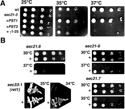
Protein trafficking is achieved by a bidirectional vesicle flow between the various compartments of the eukaryotic cell. COPII coated vesicles mediate anterograde protein transport from the endoplasmic reticulum to the Golgi apparatus, whereas retrograde Golgi-to-endoplasmic reticulum vesicles use the COPI coat. Inactivation of COPI vesicle formation in conditional sec21 (gamma-COP) mutants rapidly blocks transport of certain proteins along the early secretory pathway. We have identified the integral membrane protein Mst27p as a strong suppressor of sec21-3 and ret1-1 mutants. A C-terminal KKXX motif of Mst27p that allows direct binding to the COPI complex is crucial for its suppression ability. Mst27p and its homolog Yar033w (Mst28p) are part of the same complex. Both proteins contain cytoplasmic exposed C termini that have the ability to interact directly with COPI and COPII coat complexes. Site-specific mutations of the COPI binding domain abolished suppression of the sec21 mutants. Our results indicate that overexpression of MST27 provides an increased number of coat binding sites on membranes of the early secretory pathway and thereby promotes vesicle formation. As a consequence, the amount of cargo that can bind COPI might be important for the regulation of the vesicle flow in the early secretory pathway.


Figures

Figure 1.
Figure 1.
MST27 is a multicopy suppressor of sec21-3. Cells were grown at 25°C to an OD600 of ∼0.5. Three microliters of these cultures and 10-fold dilutions were spotted and incubated for 2 d at indicated temperatures. (A) From top to bottom, sec21wt (wt) and sec21-3 cells both transformed with an empty YEp24 plasmid, sec21-3 carrying either MST27, or PRM8 in the YEp24 vector or with the originally isolated γ1-25 plasmid. The γ1-25-harboring suppressor allows growth at 35°C, and overexpression of Mst27p enables the sec21-3 mutant to grow until 37°C. (B) The presence of γ1-25 (+) allows growth of several other γ-COP (sec21) mutants and of the α-COP/ret1 mutant sec33-1 at otherwise restrictive temperatures.
Figure 2.
Figure 2.
MST27 and PRM8 are members of a curious protein family. (A) Sequence alignment of Mst27p with its closest homologs in yeast (Mst28p, Prm8p, and Prm9p). (B) Family tree of a part of the DUP domain containing proteins, modified from PFAM database (Sonnhammer et al., 1998). (C) Predicted topologies of Mst27/28p and Prm8p. The numbers refer to amino acids as counted from the N terminus.
Figure 3.
Figure 3.
Mst27p and Mst28p as well as Prm8 and Prm9 form complexes. (A) Gel filtration analysis of the Mst27/28p and Prm8/9p complexes. The expression of myc-Mst27p or myc-Prm8p was induced upon the addition of galactose to the medium. An octylglucoside lysate from the different strains was separated in a gel filtration column and the fractions analyzed by dot immunoblot using an anti-myc antibody. (B) Determination of components of the Mst27/28p and the Prm8/9p complexes. Large-scale immunoprecipitation with the overexpressing strains for myc-Mst27p, myc-Prm8p, and a wild type were performed and separated on a large SDS gel and stained with Coomassie Blue. Specific bands were cut out and analyzed by MALDI-TOF.
Figure 4.
Figure 4.
Mst27p cycles between ER and Golgi, whereas Prm8p remains in the ER. Immunofluorescence of strains HHY217 (expressing myc-Mst27p under GAL10 promotor control) and HHY218 (expressing myc-Prm8p under GAL10 promotor control). The expression of the myc-tagged proteins was induced overnight. Rapamycin was added to the cultures for up to 6 h. The cells were processed for immunofluorescence with an anti-myc antibody and anti-mouse antibodies coupled to CY3. The DNA was visualized with 4,6-diamidino-2-phenylindole.
Figure 5.
Figure 5.
Mst27p and Mst28p bind to COPI and COPII proteins, and the C-terminal KKXX motif is required for the suppression of sec21-3. (A) The KKXX motif in Mst27p is responsible for coatomer binding. GST-fusion proteins of Mst27p, Mst27p-AAXX, and Prm8p were expressed in E. coli. The proteins were immobilized onto glutathione agarose beads and incubated with cytosol (1× cytosol, 1.25 mg/ml; 2× cytosol, 2.5 mg/ml) where indicated. After washing of the beads, the retained proteins were eluted with Laemmli buffer and analyzed by SDS-PAGE and Coomassie Blue staining. (B) GST-Mst27p-AAXX and GST-Prm8p do not bind to coatomer. Samples were treated as in A but the analysis was performed by immunoblot with antibodies directed against the coatomer complex. (C) Msts and Prm8p recruit Sec23/24p complex in a Sar1p-independent manner. The GST-fusion proteins and GST were immobilized onto glutathione agarose beads and incubated with Sar1p and Sec23/24p complex as indicated. The proteins bound to the beads were analyzed by immunoblot with anti-Sec24p antibodies. (D) Overexpression of Emp24p but not of Mst27-AAXX also suppresses the sec21-3 mutant. SEC21wt or sec21-3 cells containing an empty YEp24 vector, or sec21-3 cells overexpressing Mst27p, Mst27p-AAXX, Wbp1p, or Emp24p were grown at 25°CtoanOD600 of ∼0.5. Three microliters of these cultures and 10-fold dilutions were spotted and incubated at indicated temperatures for 2 d.
Figure 6.
Figure 6.
The γ1-25 plasmid suppresses the secretion defects in the sec21-3 mutant. Wild-type (wt) or sec21-3 mutant cells without or with the γ1-25 plasmid (+γ1-25) were grown at 25°C and shifted to 37°C for 5 min. The cells were pulse-labeled with tran35S-label for 3 min and chased at 37°C for 0–40 min as indicated. The cells were lysed and the extract used for immunoprecipitations with antiserum specific for α-factor (left) or CPY (right). The different maturation forms of CPY are indicated (p1, ER form; p2, Golgi form; m, mature in the vacuole). (B) Microsomal membranes were isolated from these cells and tested for in vitro packaging of α-factor and Sec22p. After translocation of radiolabeled α-factor, the membranes were incubated at 20°C with 8 μg/ml COPII components for the times indicated. The amounts of 35S-gpαF and Sec22p in the vesicles were quantified.
Figure 7.
Figure 7.
MST27 facilitates vesicle formation from sec21-3 membranes. (A) MST27 rescues budding defects in the presence of sec21-3 cytosol. Semi-intact cells were prepared from sec21-3 strains expressing either nothing, MST27, PRM8, or MST27-AAXX from a 2 μ plasmid. These semi-intact cells were incubated with either buffer (lanes 2, 5, 8, and 11), sec21-3 cytosol (lanes 3, 6, 9, and 12) or wild-type cytosol (lanes 4, 7, 10, and 13) for 30 min at 30°C. Vesicles released into the supernatant of a medium-speed centrifugation were concentrated by ultracentrifugation and analyzed by immunoblot. In lane 1, 10% of the total was loaded. (B) MST27 increases the release of COPI vesicles. Semi-intact cells were incubated with COPI or COPII components or wild-type cytosol for 30 min at 30°C. The vesicle formation was analyzed as described in A. (C) MST27 influences cargo uptake at the Golgi membranes. Golgi membranes were prepared from sec21-3 strains expressing either nothing, MST27, or MST27-AAXX from a 2 μ plasmid. The Golgi membranes were incubated with coatomer, GTPγS, and Arf1p for 30 min at 30°C as indicated. Membranes were floated through a sucrose cushion, resolved by SDS-PAGE and analyzed by immunoblot.
Figure 8.
Figure 8.
Co-overexpression of FLAG-Mst27p and myc-Prm8p leads to an increase in cell size and large vacuoles. Nomarski pictures of yeast cells. Cells were grown overnight in YP-galactose to induce the expression of FLAG-Mst27p, myc-Prm8p, or myc-Mst27p. The cells were subsequently observed under a light microscope with differential interference contrast. Wild type (A), HHY217 (GAL10-myc-Mst27p) (B), HHY218 (GAL10-myc-Prm8p) (C), YAS314 (HHY217 + 2 μ myc-Sec20p) (D), YAS315 (HHY218 + 2 μ myc-Sec20p) (E), and YAS254 (HHY218 + GAL1-FLAG-Mst27p (F). The bar in F represents 5 μm. The magnification in A–E is the same as in F.
Figure 9.
Figure 9.
Electron microscopy of the strains YPH500, HHY218, and YAS254. Cells were grown under the same conditions than in Figure 8 and prepared for electron microscopy. Wild type (A), HHY218 (B and C), and YAS254 (D–F). The magnification in A–C, E, and F is the same as in G. The bars in D and G represent 1.87 μm and 685 nm, respectively.
Figure 10.
Figure 10.
Co-overexpression of Mst27p and Prm8p does not result in the accumulation of the Mst27p or Prm8p complexes in the vacuole. Expression of Mst27p and Prm8p was induced overnight in strain YAS254. Cells were harvested and prepared for immunofluorescence. (A) Mst27p was visualized with an anti-FLAG antibody and CY3 coupled secondary antibodies. (C) Prm8p was stained with anti-myc and CY3 coupled secondary antibodies. (B and D) The DNA was stained with 4,6-diamidino-2-phenylindole.

Similar articles

Cited by

References

    1. Aoe, T., Cukierman, E., Lee, A., Cassel, D., Peters, P.J., and Hsu, V.W. (1997). The KDEL receptor, ERD2, regulates intracellular traffic by recruiting a GTPase-activating protein for ARF1. EMBO J. 16, 7305–7316. - PMC - PubMed
    1. Aridor, M., and Traub, L.M. (2002). Cargo selection in vesicular transport: the making and breaking of a coat. Traffic 3, 537–546. - PubMed
    1. Arnott, D., O'Connell, K.L., King, K.L., and Stults, J.T. (1998). An integrated approach to proteome analysis: identification of proteins associated with cardiac hypertrophy. Anal. Biochem. 258, 1–18. - PubMed
    1. Baker, D., Hicke, L., Rexach, M., Schleyer, M., and Schekman, R. (1988). Reconstitution of SEC gene product-dependent intercompartmental protein transport. Cell 54, 335–344. - PubMed
    1. Barlowe, C., Orci, L., Yeung, T., Hosobuchi, M., Hamamoto, S., Salama, N., Rexach, M.F., Ravazzola, M., Amherdt, M., and Schekman, R. (1994). COPII: a membrane coat formed by Sec proteins that drive vesicle budding from the endoplasmic reticulum. Cell 77, 895–907. - PubMed

Publication types

MeSH terms

Substances