. 2003 Aug;14(8):3192-207.
doi: 10.1091/mbc.e03-03-0166. Epub 2003 May 3.

Hypoxia regulates assembly of cilia in suppressors of Tetrahymena lacking an intraflagellar transport subunit gene

Affiliations

Hypoxia regulates assembly of cilia in suppressors of Tetrahymena lacking an intraflagellar transport subunit gene

Jason M Brown et al. Mol Biol Cell. 2003 Aug.

Abstract

We cloned a Tetrahymena thermophila gene, IFT52, encoding a homolog of the Chlamydomonas intraflagellar transport protein, IFT52. Disruption of IFT52 led to loss of cilia and incomplete cytokinesis, a phenotype indistinguishable from that of mutants lacking kinesin-II, a known ciliary assembly transporter. The cytokinesis failures seem to result from lack of cell movement rather than from direct involvement of ciliary assembly pathway components in cytokinesis. Spontaneous partial suppressors of the IFT52 null mutants occurred, which assembled cilia at high cell density and resorbed cilia at low cell density. The stimulating effect of high cell density on cilia formation is based on the creation of pericellular hypoxia. Thus, at least under certain conditions, ciliary assembly is affected by an extracellular signal and the Ift52p function may be integrated into signaling pathways that regulate ciliogenesis.


Figures

Figure 1.
Figure 1.
Sequence analysis and disruption of IFT52. GenBank accession numbers for sequence data used in comparisons are indicated in parentheses. (A) Ift52p (AY071864) is homologous to the known IFT proteins C. elegans OSM-6 (CAA03975) and Chlamydomonas IFT52 (AAL12162), as well as mouse NGD5 (AAA96241), human CGI53 (NP_057088), and Drosophila CG9595 (AAF52401). Black shading indicates 50% or greater identity at a given position and gray shading indicates similarity. (B and C) Southern blot analysis of germline rft1::neo2 transformants. (B) Left, Southern blot of total genomic DNA digested with EcoRI and Bsu36I and probed with a radiolabeled 3.4-kb genomic HindIII fragment of IFT52. Right, diagram of the IFT52 locus. Lane 1, control strain; lane 2, germline transformant. Arrow in right panel, site susceptible to EcoRI star activity 1.2 kb upstream of the 3′ EcoRI site. The WT locus gives fragments of 1.3 and 4.5 kb. A knockout is expected to yield a single 7.2-kb fragment. The 4.5-kb band is present in both lanes, indicating that the germline transformant is heterozygous for IFT52/ift52::neo2. Fragments generated by EcoRI star activity are indicated with asterisks. A 5.8-kb fragment likely generated by digestion of DNA with only EcoRI is indicated by δ. (C) Left, Southern blot of total genomic DNA digested with PstI and XbaI and probed with the same fragment as in B. Right, diagram of IFT52 locus. Lanes are as in B. The WT locus gives fragments of 2.0 and 5.2 kb. A knockout is predicted to give 0.6-, 2.8-, and 5.2-kb fragments. Only the fragments larger than 0.6 kb are visible. Due to heterozygosity, the 2.0-kb fragment is present in the knockout strain. Restriction sites: E, EcoRI; H, HinDIII; B, Bsu36I; P, PstI; X, XbaI.
Figure 2.
Figure 2.
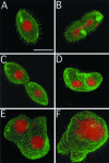
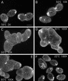
Cytological analysis of IFT52Δ cells. The phenotype was induced by conjugation of knockout heterokaryon strains UG7G5 and UG7G6. Conjugation progeny were grown in MEPPA and stained with anti-tubulin antibodies (SG) and propidium iodide. Conjugation progeny were prepared for confocal microscopy 30–31 h after isolation of conjugating pairs. (A and B) Control cells before and during division, respectively. (C) IFT52Δ mutant cell nearing the end of cytokinesis. (D) Mutant cell in an early stage after the cytokinesis failure. Two MACs are evident. (E and F) Mutants after additional cytokinesis failures forming “monster” cells. Bar, 25 μm.
Figure 3.
Figure 3.
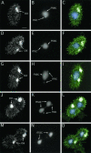
Localization of Ift52p-GFP. IFT52Δ cells rescued with a BTU1-MTT-IFT52-GFP-BTU1 fragment (see MATERIALS AND METHODS) were grown in SPP medium in the absence of cadmium. Cells were fixed and stained for confocal microscopy with polyclonal anti-GFP antibodies and DAPI. Left column, anti-GFP signal; middle column, DAPI signal; right column, merged images. (A–C) Interphase cell. Ift52p-GFP is primarily in cilia. (D–F) Cell in very early stage of micronuclear division. Posterior oral apparatus has short cilia in which Ift52p-GFP is highly concentrated. (G–I) Early stage of macronuclear elongation showing a zone of intense ciliary anti-GFP labeling immediately posterior to the developing cleavage furrow. (J–O) Later stages of macronuclear division and cleavage furrow ingression showing ciliary labeling but no accumulation in the cleavage furrow or cell body. oa, oral apparatus; mac, macronucleus; mic, micronucleus; Bar, 10 μm.
Figure 4.
Figure 4.
IFT52Δ cells were grown with (A) or without (B) shaking in MEPPA medium. After 48 h, cells were prepared for immunofluorescence microscopy as in Figure 2. Shown are merged grayscale images of the anti-tubulin and propidium iodide signals. Bar, 50 μm.
Figure 5.
Figure 5.
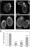
Cytological analysis of suppressor strains. Cells were grown in MEPP medium and prepared for confocal imaging by staining with anti-tubulin antibodies as in Figure 2. (A) Wild-type strain. (B) IFT52Δ10. (C) IFT52Δsm1. (D) IFT52Δmov1. (E) Quantitation of mean ciliary length (± SE of the mean) by using the measurement macro of Scion Image software. P values above each category were generated with a paired two-tailed t test with the following null hypotheses: WT = IFT52 null; IFT52 null = IFT52 sm; IFT52sm = IFT52 mov. Bar, 25 μm.
Figure 6.
Figure 6.
Electron microscopic analysis of suppressor strains. Cells prepared for EM were isolated from cultures actively growing in MEPP. (A and B) Longitudinal sections through IFT52Δ10 basal bodies. (B) A short cilium lacking a central pair is evident. (C and D) Longitudinal sections through IFT52Δsm1 basal bodies and short cilia. Some cilia in this strain have a central pair (D), whereas some do not (C). (E) Cross-sections through several IFT52Δsm1 cilia showing different degrees of completeness of central pairs. (F) Longitudinal, cross, and oblique sections through IFT52Δmov1 cilia showing that cilia are longer than in IFT52Δ10 and IFT52ΔSm1 cells and most contain central pairs. Bars, A–E, 0.25 μm; F, 1 μm.
Figure 7.
Figure 7.
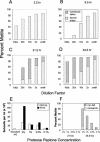
High cell density but not slow growth rate leads to ciliation in IFT52Δsm1 cells. (A–D) Exponentially growing (shaken) cells (3–5 × 10⁵/ml) were washed and resuspended in fresh MEPPA at final dilutions of 0× (undil), 2×, 10×, 20×, and 100×. Cells were maintained at 30°C without shaking, and live cells were scored on an inverted microscope for presence or absence of motility and for gross evidence of cytokinesis defects or normal cell shape. Shown are 3.5 h (A), 9.5 h (B), 21.5 h (C), and 33.5 h (D) after dilution. (E and F) IFT52Δsm1 cells were diluted to ∼3 × 10³/ml in modified MEPPA media containing varying concentrations of proteose peptone. (E) Growth rate decreased in media containing <2% proteose peptone. White bars, inoculum at t = 0 and dilutions at 20.5 h postdilution; black bars, dilutions at 44 h postdilution. (F) Cells from cultures in E were scored for motility and cytokinesis defects as in A–D.
Figure 8.
Figure 8.
Cytological analysis of IFT52Δsm1 dilution series and temperature shift experiment. Cells grown in MEPPA were washed in fresh media and resuspended at 3 × 10⁵/ml. These cells were then diluted 2× (1.5 × 105/ml) (A), 10× (3 × 104/ml) (B), 50× (6 × 103/ml) (C), and 100× (3 × 103/ml) (D and E). (F) Cells diluted 100× as in D and E were grown at 22°. After 21.5 h, cells were fixed and stained as in Figure 1.
Figure 9.
Figure 9.
Effect of shaking and mixing with other strains on ciliogenesis in IFT52Δsm suppressors. (A) IFT52Δsm1 cells were diluted to 3 × 10³/ml and grown at 22°C with or without gentle shaking or were diluted to 3 × 10⁴/ml and grown at 30°C with or without gentle shaking. Cells were scored for motility by using a 10× objective on an inverted microscope. P values were generated for comparison of proportions using a two-proportion z-test with H0: shaken = nonshaken and Ha: shaken ≠ nonshaken (n = 62 for unshaken at 30 and 22°C, 50 for shaken at 30°C, and 70 for shaken at 22°C). (B) Representative IFT52Δsm1 cells 24 h after dilution to 3 × 10⁴/ml and growth without shaking. (C) Representative IFT52Δsm1 cells 24 h after dilution to 3 × 10⁴/ml and growth with gentle shaking. (D) IFT52Δsm1or IFT52Δ10 cells were diluted to 3 × 10³/ml and incubated at 30°C without shaking and either with or without 7.5 × 10⁴ WT cells/ml. Cells were prepared for immunofluorescence as in Figure 2. Cilia lengths (± SE of the mean) were measured on single confocal sections (ncells = 20 for IFT52Δsm1 and ncells = 10 for IFT52Δ10). Numbers of individual cilia measured were 1187 for sm, 1058 for sm + WT, 126 for null, and 136 for null +WT). Indicated P values are from paired two-tailed t tests with H0: sm = sm +WT and H0: null = null +WT.
Figure 10.
Figure 10.
Hypoxia regulates assembly of cilia in IFT52Δsm suppressors. (A) IFT52Δsm7 cells were prepared at a high density of 3 × 10⁵ cell/ml and grown without shaking in either 1- or 20-μl drops or in the 10-ml volume on a 10-cm Petri plates for 20 h and scored for motility. There is an inverse relationship between the culture volume and extent of motility. (B) IFT52Δsm7 cells were suspended at the initial density of 3 × 10⁴ cells/ml and grown in varying volumes in the 250-ml Erlenmayer flasks with shaking (100 rpm). The cultures were scored for the presence of motile cells after 18 h. As the culture volume increases, the fraction of motile cells also increases. (C) IFT52Δsm7 cells were prepared at the density of 1.5 × 10⁵ cells/ml in bottles with airproof closures and subjected to a brief stream of either nitrogen or oxygen gas, or untreated (air) and left for 9.5-h incubation either shaken (130 rpm) or not at 30°C. Note high level of motility in the sample in which hypoxic conditions were created with a stream of nitrogen as well as in the normoxic sample left unshaken. Exposing cells to oxygen had an inhibitory effect on cell motility, even compared with the normoxic cells subjected to shaking. (D) Imunofluorescence image of RFTΔsm7 cells that regained motility after creation of hypoxia with a stream of nitrogen.

References

    1. Austerberry, C.F., and Yao, M.-C. (1988). Sequence structures of two developmentally regulated, alternative DNA deletion junctions in Tetrahymena thermophila. Mol. Cell. Biol. 8, 3947–3950. - PMC - PubMed
    1. Brazelton, W.J., Amundsen, C.D., Silflow, C.D., and Lefebvre, P.A. (2001). The bld1 mutation identifies the Chlamydomonas osm-6 homolog as a gene required for flagellar assembly. Curr. Biol. 11, 1591–1594. - PubMed
    1. Brown, J.M., Hardin, C., and Gaertig, J. (1999a). Rotokinesis, a novel phenomenon of cell locomotion-assisted cytokinesis in the ciliate Tetrahymena thermophila. Int. Cell Biol. Rep. 23, 841–848. - PubMed
    1. Brown, J.M., Marsala, C., Kosoy, R., and Gaertig, J. (1999b). Kinesin-II is preferentially targeted to assembling cilia and is required for ciliogenesis and normal cytokinesis in Tetrahymena. Mol. Biol. Cell 10, 3081–3096. - PMC - PubMed
    1. Cassidy-Hanley, D., Bowen, J., Lee, J., Cole, E.S., VerPlank, L.A., Gaertig, J., Gorovsky, M.A., and Bruns, P.J. (1997). Germline and somatic transformation of mating Tetrahymena thermophila by particle bombardment. Genetics 146, 135–147. - PMC - PubMed

Publication types

LinkOut - more resources