Comparative Study
. 2003 Aug;14(8):3305-24.
doi: 10.1091/mbc.e02-11-0714. Epub 2003 May 3.

Distinct mechanisms of agonist-induced endocytosis for human chemokine receptors CCR5 and CXCR4

Affiliations
Comparative Study

Distinct mechanisms of agonist-induced endocytosis for human chemokine receptors CCR5 and CXCR4

Sundararajan Venkatesan et al. Mol Biol Cell. 2003 Aug.

Abstract

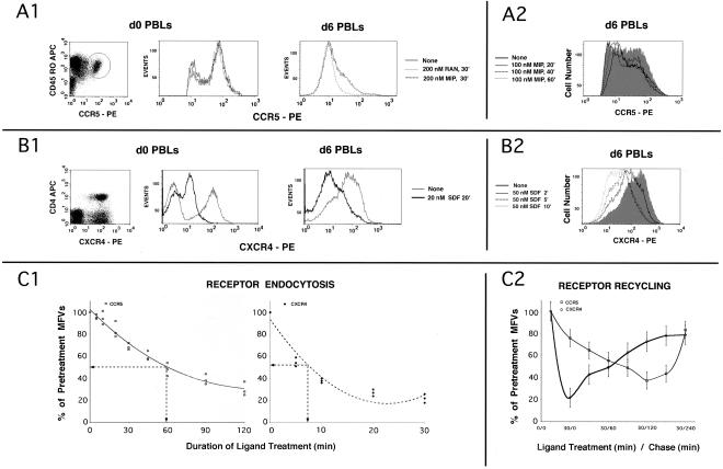
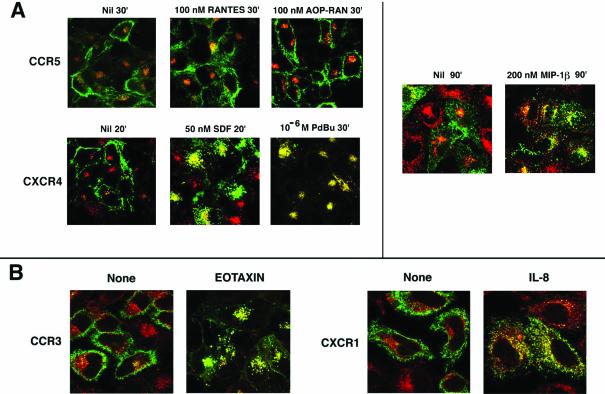
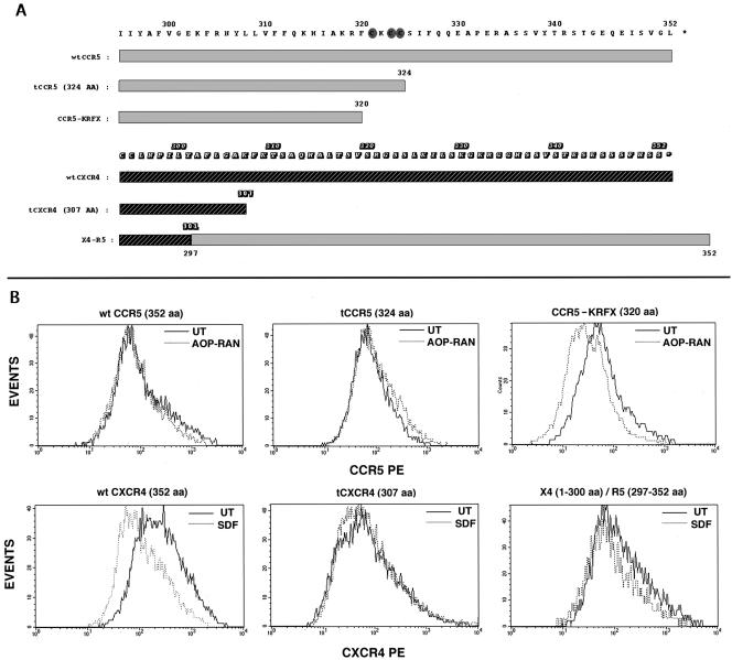
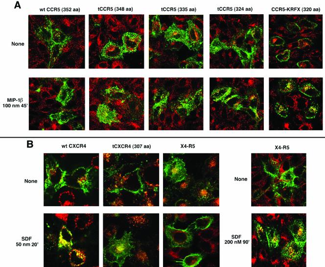
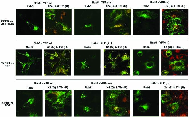
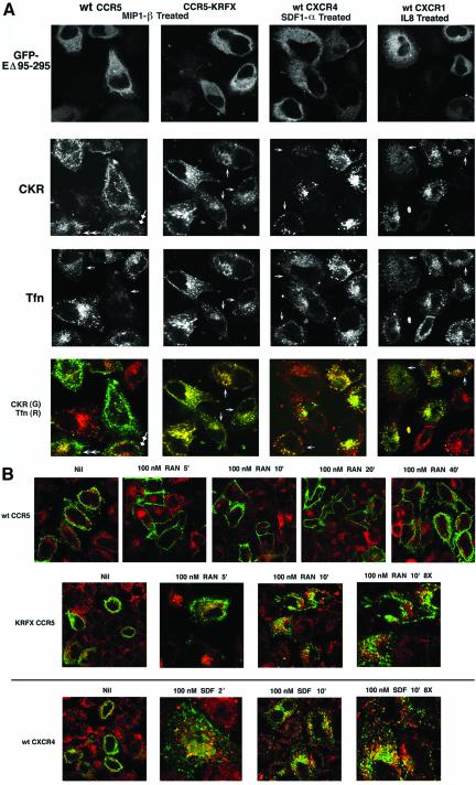
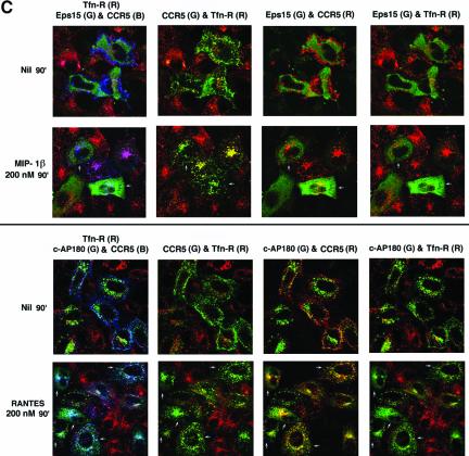
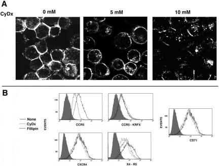
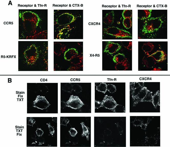
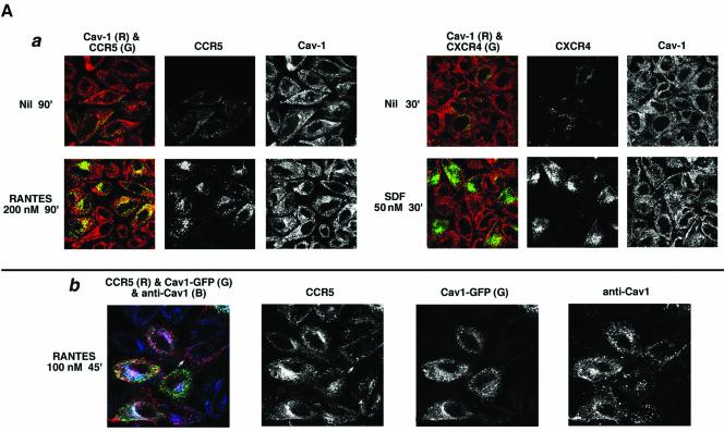
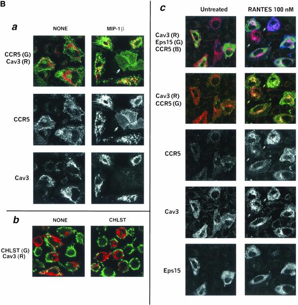
Desensitization of the chemokine receptors, a large class of G protein-coupled receptors, is mediated in part by agonist-driven receptor endocytosis. However, the exact pathways have not been fully defined. Here we demonstrate that the rate of ligand-induced endocytosis of CCR5 in leukocytes and expression systems is significantly slower than that of CXCR4 and requires prolonged agonist treatment, suggesting that these two receptors use distinct mechanisms. We show that the C-terminal domain of CCR5 is the determinant of its slow endocytosis phenotype. When the C-tail of CXCR4 was exchanged for that of CCR5, the resulting CXCR4-CCR5 (X4-R5) chimera displayed a CCR5-like trafficking phenotype. We found that the palmitoylated cysteine residues in this domain anchor CCR5 to plasma membrane rafts. CXCR4 and a C-terminally truncated CCR5 mutant (CCR5-KRFX) lacking these cysteines are not raft associated and are endocytosed by a clathrin-dependent pathway. Genetic inhibition of clathrin-mediated endocytosis demonstrated that a significant fraction of ligand-occupied CCR5 trafficked by clathrin-independent routes into caveolin-containing vesicular structures. Thus, the palmitoylated C-tail of CCR5 is the major determinant of its raft association and endocytic itineraries, differentiating it from CXCR4 and other chemokine receptors. This novel feature of CCR5 may modulate its signaling potential and could explain its preferential use by HIV for person-to-person transmission of disease.


Figures

Figure 1.
Figure 1.
(A and B) Kinetics of agonist-induced internalization of CCR5 and CXCR4, respectively, in PBLs; (C) CKR internalization and recycling in expression systems. (A1) d0 PBLs represent CD4+ T cells. Bivariate analysis of the CD4+ subset stained with CCR5 mAb 3A9-PE and CD45RO-APC is shown on the left. CCR5+ subset is encircled. Cells were stimulated with RANTES (RAN), MIP-1β (MIP) or untreated and the overlaid FACS histograms of the CCR5+ subset are shown immediately to the right. d6 PBLs are CD3 positive. Overlaid CCR5 histogram profiles of stimulated and untreated d6 PBLS are on the far right. (A2) Kinetics of MIP-1β induced CCR5 downmodulation. Overlaid FACS histograms of cells treated for various times or untreated (shaded graph) are shown. CCR5 gating parameters were adjusted to exclude low-expressers (cutoff value of ∼0.5 log). (B1) Bivariate analysis of CD3+ subset stained with CXCR4 mAb 12G5-PE; CD4-APC is on the left. CXCR4 histograms of d0 and d6 PBLs treated with SDF-1α or untreated are shown in the next two panels. (B2) Kinetics of CXCR4 downmodulation by SDF-1α. CXCR4 staining profiles of cells treated for various times or untreated (shaded graph) are shown. (C1) Receptor internalization using a HEK293 cell line expressing CCR5 and a cloned HeLa-CD4 line expressing high levels of CXCR4. Cells were stimulated with the respective chemokines (AOP-RANTES at 200 nM for CCR5; 20 nM SDF-1α for CXCR4) for the indicated times, and the MFVs of different receptors were determined by FACS analysis. Data are expressed as percentage of initial MFVs as a function of duration of chemokine treatment. Data from three experiments were used to fit a polynomial curve. (C2) For receptor recycling experiments, cell lines (noted above) expressing the indicated receptor(s) were treated with protein synthesis inhibitors before and during the experiment. CCR5 was stimulated for 30 min at 100 nM of MIP-1β and CXCR4 with 50 nM SDF-1α. MFVs of receptors at various times during the experiment are plotted as percent of MFVs of pretreated cells on the ordinate (with error bars) with time of the experiment on the abscissa.
Figure 2.
Figure 2.
Agonist-mediated CKR visualized by confocal microscopy. In all cases, Tfn-R endocytosis was monitored by Tfn-TR, shown in red. (A) Ligand mediated endocytosis of CCR5 and CXCR4 using HOS cell lines (images on the left) or HeLa cell transfectants (images on the right). CCR5 and CXCR4 were visualized by APC-labeled 3A9 and 12G5 mAb, respectively. The various ligand treatments and durations are denoted at the top of each image. Results represent three or four experiments. (B) Trafficking pattern of CCR3 and CXCR1 after exposure to their cognate ligands appropriately identified. Endocytosis assay was with HeLa cell transfectants. CCR3 and CXCR1 trafficking was visualized by use of FITC-conjugated mAbs. The respective agonists were used at 100 nM for 30 min. Results represent two experiments.
Figure 3.
Figure 3.
(A) Sequence alignment of the C-tails of CCR5, CXCR4, and derivatives. C-terminal domains of CCR5 and CXCR4 and deletions are denoted by the different shaded rectangles with the numbered amino acid sequence above. The CCR5 and CXCR4 coordinates of X4-R5 are denoted by dual shaded rectangles. Shaded ovals highlight the cysteines in the palmitoylation motif of CCR5. (B) Effect of agonist treatment on the cell surface density of CKRs. HEK293-T cells cotransfected with CD8 and the respective CKRs were treated with the appropriate agonists or untreated and stained for CD8 and CKRs. Histogram profiles of CKR densities in CD8-gated populations are shown. CCR5, tCCR5, and KRFX were stained with a mixture of CD8-APC and 3A9-PE. Agonist (AOP-RAN) treatment was at 200 nM for 30 min. CXCR4 and its derivatives were stained with CD8-APC and 12G5-PE. SDF-1α treatment was at 100 nM for 20 min. Results represent three experiments.
Figure 4.
Figure 4.
Endocytic patterns of agonist-stimulated CCR5, CXCR4, and their derivatives. Tfn-TR monitored Tfn-R trafficking. The receptors are colored green and Tfn-R in red. Images representing three experiments are shown. (A) Endocytosis pattern of AOP-RANTES (100 nM, 30 min) treated or untreated transfectants expressing wt CCR5 and serial C-terminal truncations. 3A9-APC staining visualized CCR5. (B) Endocytosis pattern of SDF-1α (50 nM, 30 min) treated or untreated cells expressing wt CXCR4, tCXCR4, or X4-R5. CXCR4 was visualized by staining with 12G5-APC. Trafficking pattern of X4-R5 chimera treated with 200 nM SDF for 90 min is shown by the images on the extreme right.
Figure 5.
Figure 5.
Agonist-driven endocytosis of CKRs in the context of wt and mutant rab5 expression. HeLa cells were cotransfected with the indicated CKRs (CKR) and wt, Q79L (++), or S34N (–) versions of rab5-YFP proteins. Tfn-TR was used to monitor Tfn-R traffic. Each row shows results obtained with a particular receptor/ligand combination. In each case, RGB images were processed to display side-by-side rab5-YFP image in green and the overlaid image of CKR in green and Tfn in red. CCR5 was stimulated with AOP-RANTES (100 nM for 30 min); CXCR4 or X4-R5 with SDF-1α. 3A9-APC and CXCR4 and X4-R5 visualized CCR5 by 12G5-APC. Images represent three experiments.
Figure 6A.
Figure 6A.
Effect of dominant negative Eps15 on agonist-driven endocytosis of wt CCR5 and KRFX mutant; wt CXCR4 and CXCR1. HeLa cells were cotransfected with GFP-tagged Eps15 mutant, EΔ95/295, and the indicated chemokine receptors (CKR). Chemokine receptor trafficking was initiated by agonist treatment (MIP-1β for wt and CCR5-KRFX, SDF-1α for wt CXCR4, and IL8 for CXCR1) at 100 nM for 30 min. APC-conjugated 3A9 or 12G5 mAb was used to visualize trafficking of CCR5 and KRFX or CXCR4, respectively. CXCR1 was stained with unconjugated mAb followed by 2o staining of fixed and permeabilized cells with APC-conjugated anti-mouse IgG. TR-conjugated Tfn was used to monitor Tfn-R trafficking. Each column represents results obtained with the individual receptor/agonist combinations that are denoted at the top. RGB images were separated into individual channels corresponding to GFP-EΔ95/295, CKR, and Tfn and laid out in successive rows. The bottom panels show overlaid images corresponding to CKR (green) and Tfn (red). Arrows denote cells described in text. Photo-multiplier gains were adjusted to visualize faint vesicles labeled with Tfn or CKR. (B) Time course of colocalization of agonist-occupied chemokine receptors with clathrin vesicles. HeLa cells transfected with the indicated receptors were treated with their cognate agonists for the indicated times or left untreated. Endocytosis was evaluated by confocal microscopy. Cells were preincubated with rabbit IgG against the N-terminal peptides of CCR5 or CXCR4 at 4°C for 15 min, before agonist treatment for the indicated times. Fixed and permeabilized (in 0.1% Triton X-100) cells were stained with mAb against human clathrin heavy chain. 2o staining was with a mixture of Alexa 488– and 568–conjugated anti-rabbit and anti-mouse IgG. Nil denotes cells without agonist for the entire duration of the assay. Images were pseudocolored to show the receptors in green and clathrin in red. All images in this figure were at 2× digital zoom using a 63× objective, except for those denoted as 8×.
Figure 6C.
Figure 6C.
Endocytic trafficking patterns of ligand bound CCR5 and Tfn-R in the context of interference of clathrin pathway by the dominant negative EΔ95/295 mutant (Eps15) or the C-terminal fragment of AP180 (c-AP180). HeLa cells were cotransected with CCR5 and GFP-tagged EΔ95/295 mutant (top panel) or FLAG-tagged c-AP180 (bottom panel). TR-conjugated Tfn illuminated Tfn-R trafficking. The top and bottom rows of each panel show results obtained without or with CCR5 chemokine treatments, respectively. The left column is a composite of images showing CCR5 mAb conjugated with APC (CCR5; B), Tfn-R in red, and GFP-tagged, EΔ95/295 (Eps15; G) in the top panel or c-AP180 stained with rabbit anti-FLAG IgG followed by Alexa 568 conjugated 2o antibody in the bottom panel. Pseudocolored images representing CCR5 in green and Tfn-R in red; CCR5 in red and Eps15 mutant or c-AP180 in green; and Tfn-R in red and Eps15 mutant or c-AP180 in green are shown in the successive columns on the right. Arrows denote cells described in text.
Figure 7.
Figure 7.
Cholesterol depletion by methyl-β-cyclodextrin (CyDx) and its effect on cell surface density of CKRs. (A) Cholesterol staining of CyDx-treated HeLa cells. After treatment, cells were fixed and stained for cholesterol with fillipin (10 μg/ml) for 10 min at RT and visualized by UV laser microscopy. (B) FACS histograms of CKR expression in HeLa cell transfectants treated with CyDx (5 mM) or fillipin (10 μg/ml) or left untreated. CD4 was cotransfected in each case to monitor expression. Transfectants were stained with CD4-APC and PE-conjugated mAbs for the respective receptors, except for the KRFX mutant. Because transport of this mutant to the cell surface was quite poor, cells were stained with unconjugated 1o mAbs followed by PE-conjugated 2o antibodies. Cells were gated for CD4 staining because it was not altered by cyclodextrin or fillipin treatment. Histogram profiles of CKR densities in CD4-gated populations representing two experiments are shown. Shaded histograms represent staining with an isotype control.
Figure 8.
Figure 8.
(A) Copatching of CCR5, CXCR4, and derivatives with raft and nonraft markers. HeLa cells transfected with wt CCR5, KRFX, CXCR4, or X4-R5 were stained at 20°C for 10 min with FITC-labeled 2D7 (for CCR5 and KRFX) or 12G5 mAbs for wt and KRFX-CCR5 or CXCR4, respectively; CD71-APC followed by cross-linking with anti-mouse IgG. For copatching with CTx-B, cells were costained with Alexa 594-CTx-B and unlabeled 2D7 or 12G5 mAb at 20°C for 15 min followed by 20°C staining with Alexa 488–conjugated anti-mouse IgG. CKRs are in green, whereas Tfn-R and CTx-B are in red. Images represent three experiments. (B) Cell surface distribution of receptors after detergent extraction. For visualizing CCR5, CD4, and Tfn-R in the same background, HeLa cells were transfected with CCR5 and CD4. CXCR4 was visualized in a HeLa CD4 clone expressing high levels of CXCR4. The two different protocols are described as in MATERIALS AND METHODS. CCR5 transfectants were stained with a mixture of CD4-FITC, 3A9-APC, and CD71-APC to visualize CD4, CCR5, and Tfn-R, respectively. CXCR4 was visualized using APC-conjugated 12G5 mAb. Single-channel images from two experiments are shown.
Figure 9A.
Figure 9A.
Caveolin-1 and -3 differentially regulate agonist-dependent endocytosis of CCR5 and CXCR4. HeLa cells were transfected with the indicated chemokine receptor and treated with the indicated agonists. Cell surface–bound antibody was stripped by acid wash. Color overlays and individual channels are laid out left to right. R, red; G, green; B, blue. (a) Endocytic trafficking patterns of agonist-occupied CCR5 and CXCR4 in HeLa cells stained for endogenous caveolin-1. Receptor endocytosis was visualized by antibody feeding using APC-conjugated 1o mAbs (pseudocolored green). Caveolin-1 was detected by staining with rabbit anticaveolin-1 IgG followed by 2o staining with Alexa 488–conjugated anti-rabbit IgG (red). (b) CCR5 trafficking in HeLa cells overexpressing caveolin1-GFP. Agonist treatment was restricted to 45 min at 100 nM to limit CCR5 endocytosis. CCR5 trafficking was visualized by feeding unconjugated 3A9 mAb. Internalized antibody was visualized by staining with Alexa 568–conjugated anti-mouse IgG. Fluorescence from exogenous Cav1-GFP is in green. Both endogenous and plasmid-expressed caveoin-1 were detected by staining with rabbit IgG against caveolin-1 followed by staining with Alexa 647–labeled anti-rabbit IgG as the 2o reagent. Note that Cav1-GFP fluorescence and Cav1 antibody staining do not overlap completely, presumably because the epitope recognized by rabbit antibody is not exposed on all caveolin molecules.
Figure 9B.
Figure 9B.
Overexpression of caveolin-3 (Cav3). Agonist-treated CCR5 is internalized into caveolin-positive vesicles by largely clathrin-independent pathways, and these cells display selective depletion of plasma membrane cholesterol. Antibody feeding with APC-conjugated 3A9 mAbs monitored the trafficking of CCR5 induced by the indicated agonists at 100 nm for 30 min. HA-tagged Cav-3 (Cav3) was detected by staining with biotin-conjugated anti-HA mAb, followed by Alexa 488– (a) or TR-(c) labeled neutravidin. Images reflect results of three experiments. R, red; G, green; B, blue. (a) Composite images of CCR5 (green) and Cav3 (red) are shown at the top, with the monochromatic images of CCR5 and Cav3 below. (b) Staining for cell surface cholesterol in Cav3 transfectants. Cells were fixed and stained for cholesterol (CHLST, green) by fillipin (10 μg/ml) and then permeabilized and stained for Cav3 (red). (c) Eps 15 mutant does not eliminate Cav3-enhanced endocytosis of ligand-occupied CCR5. HeLa cells were cotransfected with HA-tagged Cav3 (Cav3), GFP-tagged mutant EΔ95/295 of Eps15 (Eps15), and CCR5. Receptor endocytosis assay was as above except that cell surface–bound antibody was stripped by acid wash. Trichromatic, bichromatic, and monochromatic images are displayed from top to bottom.

Similar articles

Cited by

References

    1. Alkhatib, G., Locati, M., Kennedy, P.E., Murphy, P.M., and Berger, E.A. (1997). HIV-1 coreceptor activity of CCR5 and its inhibition by chemokines: independence from G protein signaling and importance of coreceptor downmodulation. Virology 234, 340–348. - PubMed
    1. Alonso, M.A., and Millan, J. (2001). The role of lipid rafts in signalling and membrane trafficking in T lymphocytes. J. Cell Sci. 114, 3957–3965. - PubMed
    1. Amara, A., Gall, S.L., Schwartz, O., Salamero, J., Montes, M., Loetscher, P., Baggiolini, M., Virelizier, J.L., and Arenzana-Seisdedos, F. (1997). HIV coreceptor downregulation as antiviral principle: SDF-1alpha-dependent internalization of the chemokine receptor CXCR4 contributes to inhibition of HIV replication. J. Exp. Med. 186, 139–146. - PMC - PubMed
    1. Anderson, R.G. (1998). The caveolae membrane system. Annu. Rev. Biochem. 67, 199–225. - PubMed
    1. Aramori, I., Ferguson, S.S., Bieniasz, P.D., Zhang, J., Cullen, B., and Cullen, M.G. (1997). Molecular mechanism of desensitization of the chemokine receptor CCR-5, receptor signaling and internalization are dissociable from its role as an HIV-1 co-receptor. EMBO J. 16, 4606–4616. - PMC - PubMed

Publication types

LinkOut - more resources