. 2003 Aug;14(8):3389-99.
doi: 10.1091/mbc.e02-11-0704. Epub 2003 Apr 17.

Subcellular localization and activity of multidrug resistance proteins

Affiliations

Subcellular localization and activity of multidrug resistance proteins

Asha Rajagopal et al. Mol Biol Cell. 2003 Aug.

Abstract

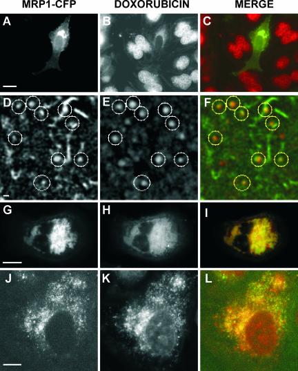
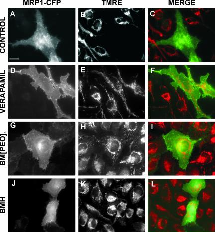
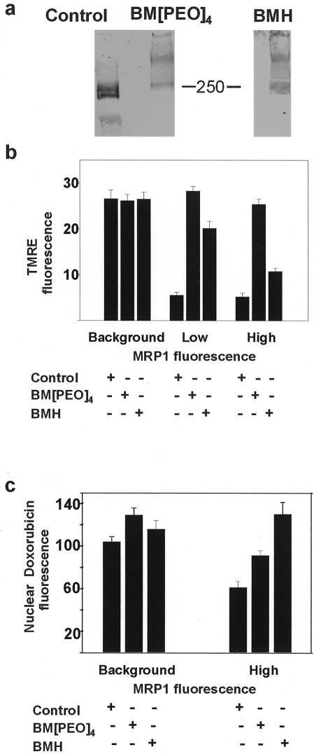
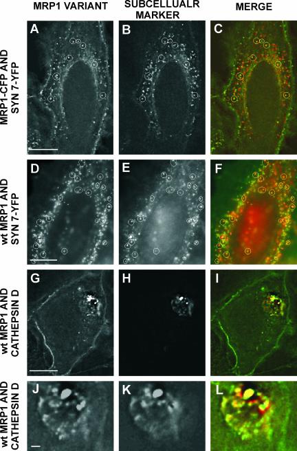
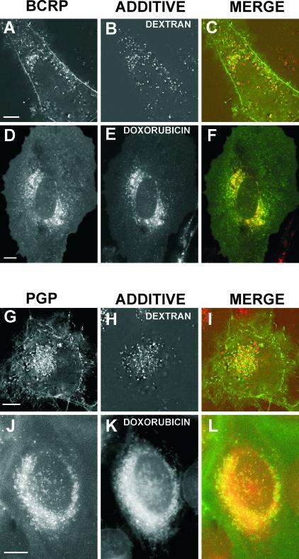
The multidrug resistance (MDR) phenotype is associated with the overexpression of members of the ATP-binding cassette family of proteins. These MDR transporters are expressed at the plasma membrane, where they are thought to reduce the cellular accumulation of toxins over time. Our data demonstrate that members of this family are also expressed in subcellular compartments where they actively sequester drugs away from their cellular targets. The multidrug resistance protein 1 (MRP1), P-glycoprotein, and the breast cancer resistance protein are each present in a perinuclear region positive for lysosomal markers. Fluorescence-activated cell sorting analysis suggests that these three drug transporters do little to reduce the cellular accumulation of the anthracycline doxorubicin. However, whereas doxorubicin enters cells expressing MDR transporters, this drug is sequestered away from the nucleus, its subcellular target, in vesicles expressing each of the three drug resistance proteins. Using a cell-impermeable inhibitor of MRP1 activity, we demonstrate that MRP1 activity on intracellular vesicles is sufficient to confer a drug resistance phenotype, whereas disruption of lysosomal pH is not. Intracellular localization and activity for MRP1 and other members of the MDR transporter family may suggest different strategies for chemotherapeutic regimens in a clinical setting.


Figures

Figure 1.
Figure 1.
The effect of MRP1-ECFP localization on the distribution of doxorubicin. (A) ECFP fluorescence reveals that a transiently transfected HeLa cell expresses MRP1 both at the plasma membrane and in a juxtanuclear region. Bar, 10 μm. (B) Doxorubicin fluorescence demonstrates that the drug likewise accumulates in a juxtanuclear region in an MRP-ECFP–expressing cell, whereas the nonexpressing cells surrounding it accumulate the drug in the nucleus. (C) The merge of MRP1-ECFP fluorescence (green) and doxorubicin fluorescence (red) shows the colocalization of doxorubicin and perinuclear localized MRP1-ECFP (yellow). (D–F) An enlarged image of the cell depicted in A–C reveals individual MRP1-ECFP–containing vesicles that also contain doxorubicin. Bar, 1 μm. (G–I) In rare instances when MRP1-ECFP aberrantly collects in the endomembrane system, doxorubicin accumulation is not juxtanuclear but is likewise dispersed. Bar, 10 μm. (J–L) A cell expressing low levels of MRP1-ECFP has little to no plasma membrane ECFP fluorescence and accumulates doxorubicin in a pattern that coincides with intracellular MRP1-ECFP. Bar, 5 μm.
Figure 2.
Figure 2.
Cross-linking MRP1-ECFP interferes with its ability to transport substrates at the plasma membrane, as assayed by TMRE accumulation. (A–C) MRP1-ECFP expression prevents the intracellular accumulation of TMRE, so that the MRP1-positive cell is not visible under TMRE fluorescence. (D–F) Inhibiting MRP1 with verapamil prevents MRP1-mediated TMRE transport, and the two MRP1-ECFP–expressing cells in this field now accumulate the drug. (G–I) Cross-linking cells with the cell-impermeable reagent BM[PEO]4 prevents MRP1-mediated TMRE transport so that the MRP1-positive cell accumulates TMRE just like its nonexpressing counterparts. (J–L) Addition of the cell permeable cross-linker BMH also inhibits MRP1 transport of TMRE. Bar, 10 μm.
Figure 3.
Figure 3.
MRP1-ECFP actively sequesters doxorubicin in internal compartments. (A–C) Use of the MRP1 inhibitor verapamil redistributes doxorubicin from the juxtanuclear region to the nucleus of all three cells expressing the protein. (D–F) Addition of the cellimpermeable cross-linker BM[PEO]4 has no effect on the subcellular distribution of doxorubicin in MRP1-ECFP–expressing cells; the drug is still to be found in juxtanuclear regions that are MRP1-ECFP positive. (G–I) Addition of the cell-permeable cross-linker BMH redistributes doxorubicin to the nucleus of MRP1-ECFP–expressing cells, much as the MRP1 inhibitor verapamil did in A–C. (J–L) Disrupting the organellar pH of cells with concanamycin A does not effect MRP1-ECFP–mediated drug sequestration; doxorubicin is still excluded from the nucleus of MRP1-expressing cells. Bar, 10 μm.
Figure 4.
Figure 4.
The effect of cross-linking on the electrophoretic mobility and transport activities of MRP1-ECFP. (A) In an immunoblot of MRP1-ECFP–transfected cell lysates, an anti-MRP1 antibody recognizes a doublet whose molecular mass migrates below the 250-kDa protein marker. However, addition of BM[PEO]4 significantly retards the mobility of MRP1-ECFP. An immunoblot of BMH-treated cells likewise reveals a changed mobility of the protein after cross-linking. (B) MRP1-ECFP activity can be quantified by relating how much TMRE a cell accumulates to how much MRP1-ECFP a cell expresses. Fluorescence functions as a reporter for both MRP1 expression and TMRE accumulation. Neither BM[PEO]4 nor BMH increases the permeability of cells to TMRE, because all cells with background MRP1 fluorescence accumulate comparable levels of TMRE. Moreover, cells treated with BM[PEO]4, regardless of the degree to which they express MRP1, accumulate as much TMRE as untreated, non-MRP1–expressing cells. (C) MRP1-ECFP activity against doxorubicin can be quantified by relating MRP1 expression to the doxorubicin fluorescence inside the nucleus. In BM[PEO]4-treated cells, MRP1-ECFP still reduces the relative amount of doxorubicin accumulated in the nucleus. For both BM[PEO]4-treated and untreated cells, MRP1-mediated reduction in nuclear fluorescence is statistically significant (P < 0.01 and p < 0.0001, respectively). However, all BMH-treated cells have similar amounts of doxorubicin in the nucleus, whether they express MRP1-ECFP or not.
Figure 5.
Figure 5.
Fluorescently labeled MRP1 and wild-type MRP1 both colocalize with lysosomal markers. (A) An MRP1-YFP–transfected cell expresses MRP1 at the plasma membrane and in intracellular vesicles (white circles). Bar, 10 μm. (B) Cotransfected with synaptotagminVII-YFP, the cell in A also expresses a lysosomal marker in intracellular vesicles (white circles). (C) The merge of the MRP1-YFP fluorescence in A (green) with the synaptotagmin fluorescence in B (red) reveals the degree of colocalization. Images are of living cells with moving vesicles; a delay of at least 1 s separates the images in time. (D–F) The degree of colocalization of wild-type MRP1 (green) and synaptotagminVII (green). Bar, 10 μm. (G–I) Localization patterns of wild-type MRP1 (green) and cathepsin D (red). Bar, 10 μm. (J–L) An enlarged image of the cell depicted in G–I reveals the degree of colocalization of the two proteins. Bar, 1 μm.
Figure 6.
Figure 6.
Pgp and BCRP colocalize with a lysosomal marker and accumulate doxorubicin in regions positive for either Pgp or BCRP. Bars, 10 μm. (A) A cell transfected with BCRP-ECFP expresses the protein at the plasma membrane and in intracellular regions. The image is a deconvolved fluorescent section of a cell. (B) Fluorescent dextrans chased into the lysosomes of the cell in A accumulate in intracellular vesicles. (C) The merge of A and B shows the degree to which BCRP-ECFP (green) colocalizes with the lysosomal marker (red). (D–F) The degree of colocalization of BCRP-ECFP in D and doxorubicin in E. (G–I) The degree to which Pgp-ECFP accumulates in intracellular vesicles G that are positive for fluorescent dextrans chased into the lysosomes H. (J–L) Localization patterns of Pgp-ECFP and doxorubicin.
Figure 7.
Figure 7.
Correlation coefficients quantify degrees of correlation for fluorescent markers. (A) Line graph plotting the correlation coefficients of MRP1-ECFP in differently treated cells. (B and C) Line graphs plotting the correlation coefficients of MRP1-ECFP and wild-type MRP1 with various subcellular markers. (D) Line graph plotting the correlation coefficients of specified ABC proteins and an additive which is either a lysosomal marker or doxorubicin.

Similar articles

Cited by

References

    1. Altan, N., Chen, Y., Schindler, M., and Simon, S.M. (1998). Defective acidification in human breast tumor cells and implications for chemotherapy. J. Exp. Med. 187, 1583–1598. - PMC - PubMed
    1. Cabrita, M.A., Hobman, T.C., Hogue, D.L., King, K.M., and Cass, C.E. (1999). Mouse transporter protein, a membrane protein that regulates cellular multidrug resistance, is localized to lysosomes. Cancer Res. 59, 4890–4897. - PubMed
    1. Chang, X.B., Hou, Y.X., and Riordan, J.R. (1997). ATPase activity of purified multidrug resistance-associated protein. J. Biol. Chem. 272, 30962–30968. - PubMed
    1. Chen, Y., Schindler, M., and Simon, S.M. (1999). A mechanism for tamoxifen-mediated inhibition of acidification. J. Biol. Chem. 274, 18364–18373. - PubMed
    1. Chen, Y., and Simon, S.M. (2000). In situ biochemical demonstration that P-glycoprotein is a drug efflux pump with broad specificity. J. Cell Biol. 148, 863–870. - PMC - PubMed

Publication types

MeSH terms

Substances