Comparative Study
. 2003 Sep;6(9):989-95.
doi: 10.1038/nn1111.

Faces and objects in macaque cerebral cortex

Affiliations
Comparative Study

Faces and objects in macaque cerebral cortex

Doris Y Tsao et al. Nat Neurosci. 2003 Sep.

Abstract

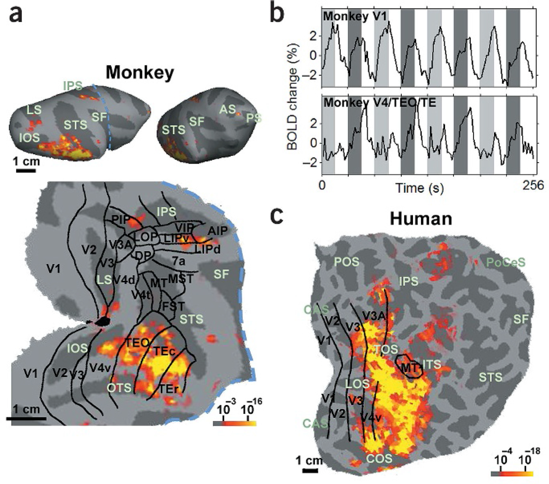
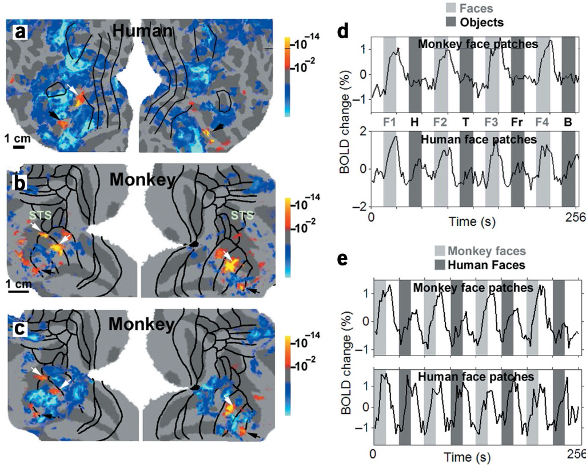
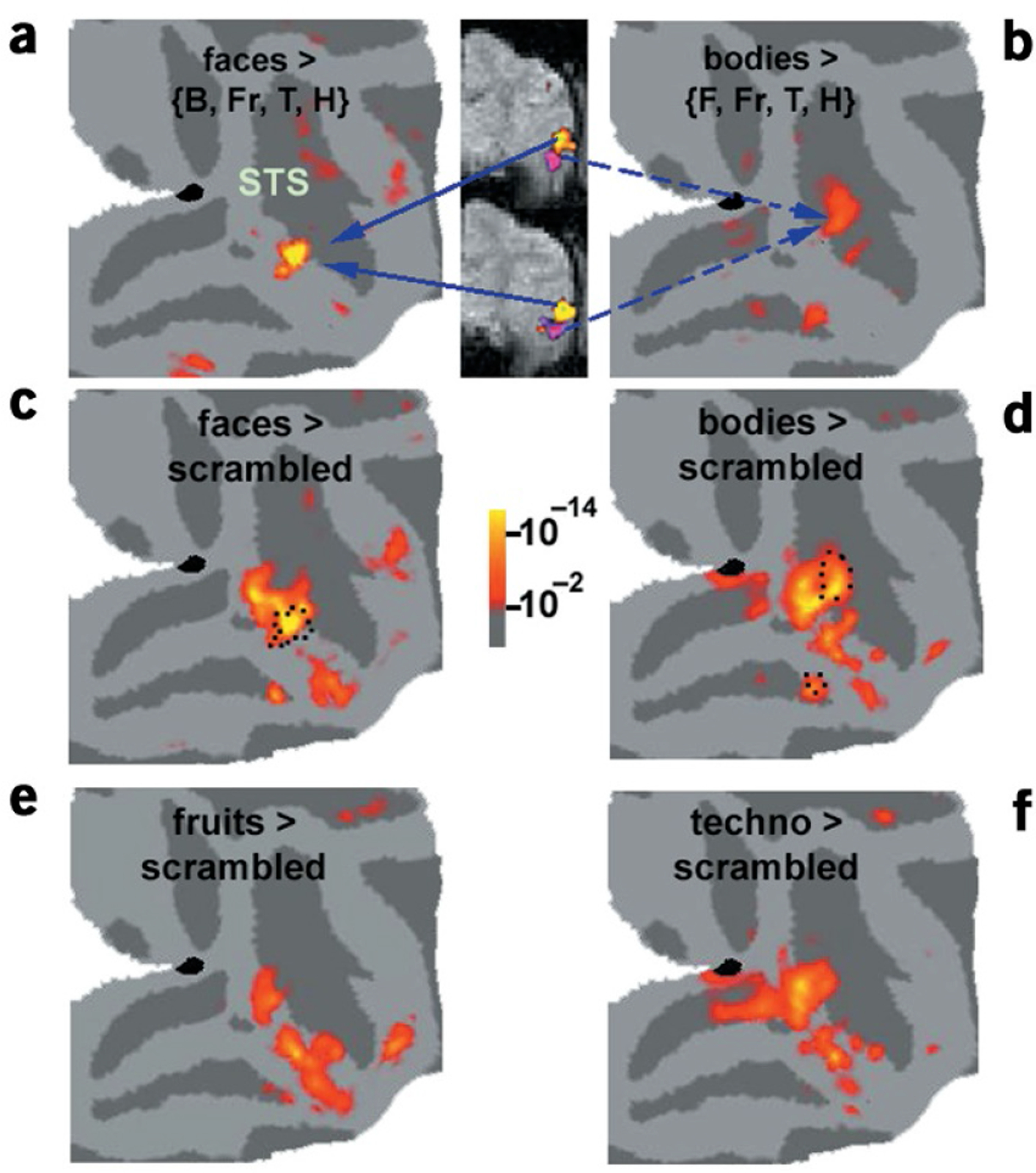
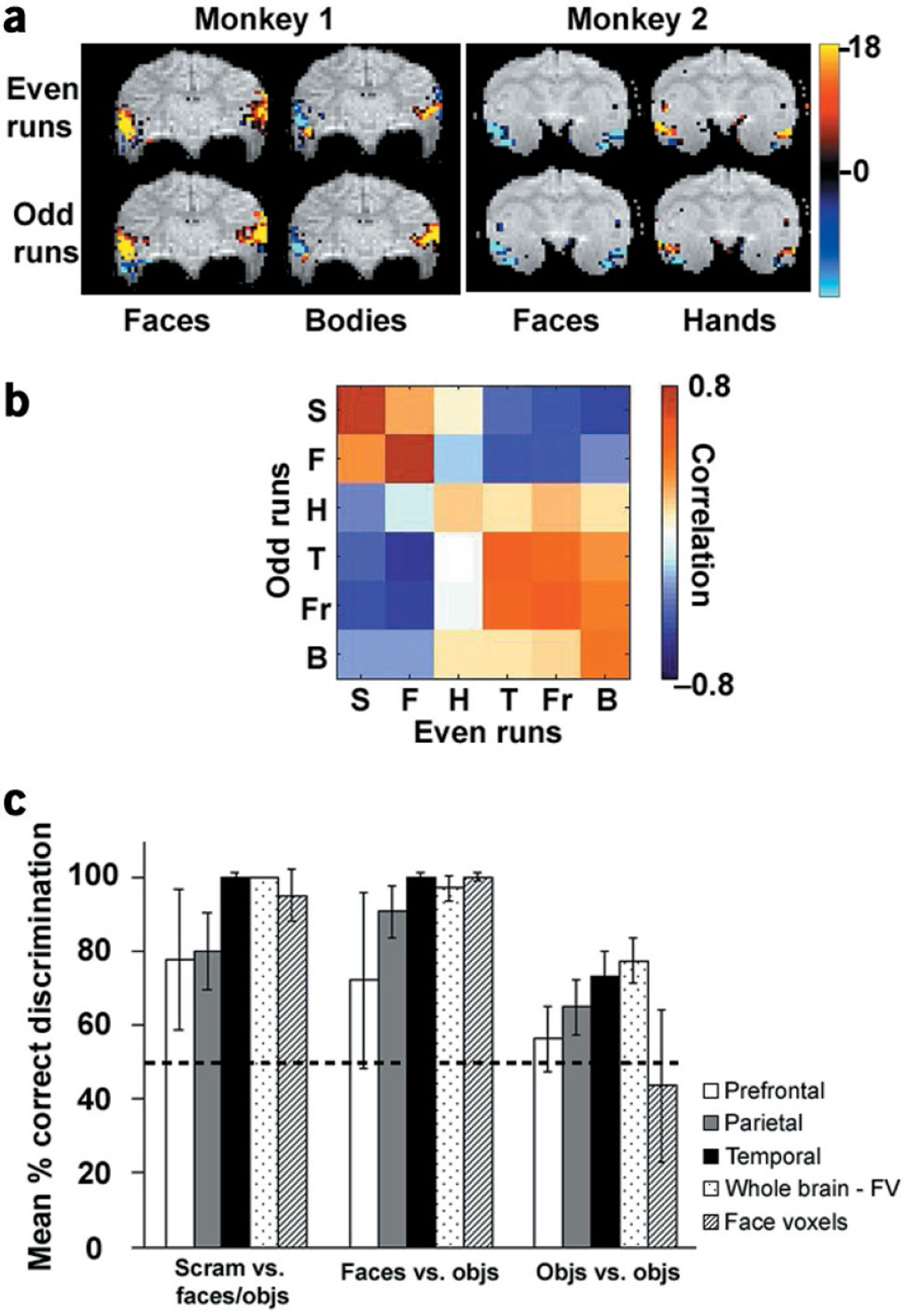
How are different object categories organized by the visual system? Current evidence indicates that monkeys and humans process object categories in fundamentally different ways. Functional magnetic resonance imaging (fMRI) studies suggest that humans have a ventral temporal face area, but such evidence is lacking in macaques. Instead, face-responsive neurons in macaques seem to be scattered throughout temporal cortex, with some relative concentration in the superior temporal sulcus (STS). Here, using fMRI in alert fixating macaque monkeys and humans, we found that macaques do have discrete face-selective patches, similar in relative size and number to face patches in humans. The face patches were embedded within a large swath of object-selective cortex extending from V4 to rostral TE. This large region responded better to pictures of intact objects compared to scrambled objects, with different object categories eliciting different patterns of activity, as in the human. Overall, our results suggest that humans and macaques share a similar brain architecture for visual object processing.


Conflict of interest statement

Competing Interests Statement

The authors declare that they have no competing financial interests.

Figures

Figure 1
Figure 1
Object-selective areas in the macaque and human. (a) Areas in the macaque brain significantly (P < 10–3) more activated by intact objects than by grid-scrambled counterparts. The activation is rendered on two different lateral views of the inflated right hemisphere (top), as well as on a flat map of the right visual cortex (bottom). The blue dashed line indicates the cut used to construct the flat map. The borders of macaque visual areas, including the Ungerleider and Desimone partitioning scheme for temporal regions (ventral V4, TEO, TEr and Tec) and the Lewis and Van Essen scheme for all other regions, were derived by warping a macaque atlas to the individual hemisphere with surface-based registration, using major sulci and gyri as landmarks. Significant activation occurred in ventral stream areas V4, TEO and TE, as well as in two additional foci—the lateral bank of the intraparietal sulcus and inferior prefrontal cortex. (b) Time courses from macaque V1 (top) and V4/TEO/TE (bottom) to alternating epochs of intact (dark gray) and scrambled (light gray) objects, separated by epochs of a blank screen (white). (c) Activity in the right hemisphere of a human subject to the same stimulus comparison. The borders of human visual areas were determined by retinotopic and functional mapping. Sulcal abbreviations: LS, lunate; IOS, inferior occipital; OTS, occipito-temporal; IPS, intraparietal; STS, superior temporal; SF, Sylvian fissure; AS, arcuate; ITS, inferior temporal; POS, parieto-occipital; CAS, calcarine; COS, collateral; TOS, transoccipital; LOS, lateral occipital; PoCeS, postcentral.
Figure 2
Figure 2
Face-selective patches in the human and macaque. (a) Patches in human visual cortex significantly (P < 10–2) more activated by faces than by non-face objects. Activation maps are rendered on flattened views of the left and right hemispheres (on left and right of panel, respectively). This subject showed FFA activity in each hemisphere (black arrows), as well as two additional face-selective patches: in the left posterior inferior temporal gyrus (white arrow) and in the right anterior superior temporal sulcus (region not covered by the right hemisphere flat map). (b) Face-selective patches in a macaque, derived using the same stimuli and analysis as for the human (see panel a in Supplementary Fig. 4 for same data in slice format). The three most significant face patches (two in the left hemisphere, one in the right hemisphere) were located in the fundus and lower bank of the STS (white arrows). Two additional face-selective patches were located bilaterally in rostral TE (black arrows). In addition, there was a bilateral patch in the STS in area TEO that was not as reliably imaged across different days and was not found for line drawings. (c) Face-selective patches in the macaque, derived using line drawings of faces and objects instead of grayscale photographs. Fiducial arrows mark the face patches in both b and c. (d) Time courses from the monkey and human face patches to face (light grey) versus non-face (dark grey) object stimuli; during white-coded epochs, the stimuli were Fourier-phase scrambled counterparts of images in the subsequent colored epoch. A different set of faces (F1–F4) and objects were presented during each of the purple- and blue-coded epochs. H, hands; T, technological objects; Fr, fruits; B, bodies. (e) Time courses from the monkey and human face patches (defined based on selectivity to human faces) to macaque (light grey) versus human (dark grey) face stimuli.
Figure 3
Figure 3
Regions of maximal and relative category selectivity. (a,b) Patches of macaque cortex maximally responsive to faces and to bodies, displayed on the same right-hemisphere map. The body and face-selective patches are also shown on two functional slices, to highlight their proximity. (c–f) Regions significantly more activated by each object category than by grid-scrambled objects. The dotted lines in c and d outline the region of absolute selectivity shown in a and b. The response patterns to the different categories were distributed and partially overlapping.
Figure 4
Figure 4
The information content of distributed activity patterns in different brain regions. (a) An exemplary slice from each of two monkeys, showing the response pattern to two different object categories during even and odd runs. (b) Color-coded matrix of correlation values between responses to each object category during even and odd runs (abbreviations as in Fig. 2; S, scrambled technological objects). Only visually activated voxels (P < 0.01) in the temporal lobe were used to compute the correlation matrix, and data were averaged across two monkeys. (c) Mean percentage pairwise correct discrimination (see Methods) and standard errors across two scan sessions in two monkeys for three different discrimination types (faces and objects versus scrambled, faces versus objects, and objects versus objects) based on visually activated voxels in the prefrontal lobe (white), parietal lobe (gray), temporal lobe (black), whole brain excluding face-selective voxels (stippled) and face-selective voxels only (striped). For each discrimination type, chance performance would be 50%. All data were averaged across two experimental sessions in two monkeys.

Similar articles

Cited by

References

    1. Ungerleider L & Mishkin M Two cortical visual systems. in Analysis of Visual Behavior (ed. Ingle D) 549–586 (MIT Press, Cambridge, Massachusetts, 1982).
    1. Logothetis N & Sheinberg D Visual object recognition. Annu. Rev. Neurosci 19, 577–621 (1996). - PubMed
    1. Gross CG, Rocha-Miranda CE & Bender DB Visual properties of neurons in inferotemporal cortex of the macaque. J. Neurophysiol 35, 96–111 (1972). - PubMed
    1. Desimone R, Albright T, Gross CG & Bruce C Stimulus-selective properties of inferior temporal neurons in the macaque. J. Neurosci 4, 2051–2062 (1984). - PMC - PubMed
    1. Perrett D, Hietanen J, Oram M & Benson P Organization and function of cells responsive to faces in the temporal cortex. Phil. Trans. R. Soc. Lond 335, 23–30 (1992). - PubMed

Publication types