

The diagnosis and management of pre-invasive breast disease: flat epithelial atypia--classification, pathologic features and clinical significance
- PMID: 12927037
- PMCID: PMC314429
- DOI: 10.1186/bcr625
The diagnosis and management of pre-invasive breast disease: flat epithelial atypia--classification, pathologic features and clinical significance
Abstract
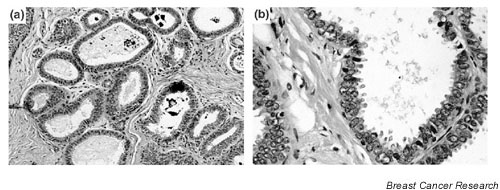
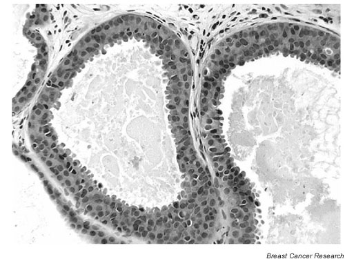
Flat epithelial atypia is a descriptive term that encompasses lesions of the breast terminal duct lobular units in which variably dilated acini are lined by one to several layers of epithelial cells, which are usually columnar in shape and which display low-grade cytologic atypia. Observational studies have suggested that at least some of these lesions may represent either a precursor of ductal carcinoma in situ (DCIS) or the earliest morphological manifestation of DCIS. In contrast, the limited available clinical follow-up data suggest that the risk of both local recurrence and progression of these lesions to invasive cancer is extremely low, supporting the notion that categorizing such lesions as 'clinging carcinoma' and managing them as if they were fully developed DCIS will result in overtreatment of many patients. Additional studies are needed to better understand the biological nature and clinical significance of these lesions.
Figures
References
-
- Tavassoli FA, Hoefler H, Rosai J, Holland R, Ellis I, Schnitt S, Lakhani SRM, Boecker W, Heywang-Kobrunner SH, Moinfar F, Peterse J. Intraductal proliferative lesions. In: Tavassoli FA, Devilee P, editor. Pathology and Genetics of Tumours of the Breast and Female Genital Organs. Lyon: IARC Press;
-
- Tsuchiya S. Atypical ductal hyperplasia, atypical lobular hyperplasia, and interpretation of a new borderline lesion. Jpn J Cancer Clin. 1998;44:548–555.
-
- Oyama T, Iijima K, Takei H, Horiguchi J, Iino Y, Nakajima T, Koerner F. Atypical cystic lobule of the breast: an early stage of low-grade ductal carcinoma in-situ. Breast Cancer. 2000;7:326–331. - PubMed
-
- Wellings SR, Jensen HM, Marcum RG. An atlas of subgross pathology of the human breast with special reference to possible precancerous lesions. J Natl Cancer Inst. 1975;55:231–273. - PubMed
-
- Azzopardi JG. Problems in Breast Pathology. Philadelphia, PA: WB Saunders. 1979. - PubMed
Publication types
MeSH terms
LinkOut - more resources
Full Text Sources
Other Literature Sources
Medical
