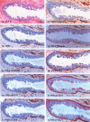
A subset of morphologically distinct mammary myoepithelial cells lacks corresponding immunophenotypic markers
- PMID: 12927046
- PMCID: PMC314436
- DOI: 10.1186/bcr635
A subset of morphologically distinct mammary myoepithelial cells lacks corresponding immunophenotypic markers
Abstract
Introduction: Immunostaining for smooth muscle actin (SMA) is commonly used to elucidate mammary myoepithelial (ME) cells, whose presence or absence is a reliable criterion for differentiating in situ and invasive carcinomas. However, some morphologically distinct ME cells fail to stain for SMA. This study intended to assess whether these SMA-negative cells also lack the expression of other ME cell markers.
Methods: Hematoxylin/eosin and SMA immunostained sections from 175 breast cancer patients were examined. Three cases were found to harbor ducts that showed morphologically distinct ME cell layers, but showed no SMA immunostaining in at least one-third of the layer or the entire layer. Eight additional consecutive sections from each case were stained for SMA, using a black chromogen, and each was then re-stained for one of eight additional markers supposed to exclusively or preferentially stain ME cells, using a red chromogen. SMA-negative ME cells were re-examined for the expression of other markers.
Results: SMA-negative ME cells in two cases also failed to display immunoreactivity for other markers, including calponin, CD10, smooth muscle myosin heavy chain, protease inhibitor 5 (maspin), Wilms' tumor-1, and cytokeratins 5, 14, and 17 (CK5, CK14, and CK17). However, in one case SMA-negative ME cells displayed immunoreactivities for maspin, CK5, CK14, and CK17. The distribution of these ME cells is independent of ductal size, length, and architecture.
Conclusions: A subset of morphologically identifiable ME cells lack the expression of nine corresponding immunophenotypic markers, suggesting that ME cells might also be subject to different normal and pathological alterations.
Figures


Similar articles
-
Phenotypic and Functional Characterization of Ductal Carcinoma In Situ-Associated Myoepithelial Cells.Clin Breast Cancer. 2015 Oct;15(5):335-42. doi: 10.1016/j.clbc.2015.01.004. Epub 2015 Jan 21. Clin Breast Cancer. 2015. PMID: 25700939
-
Availability of CD10 immunohistochemistry as a marker of breast myoepithelial cells on paraffin sections.Mod Pathol. 2002 Apr;15(4):397-405. doi: 10.1038/modpathol.3880536. Mod Pathol. 2002. PMID: 11950913
-
Dual usages of single Wilms' tumor 1 immunohistochemistry in evaluation of breast tumors: a preliminary study of 30 cases.Cancer Biomark. 2009;5(3):109-16. doi: 10.3233/CBM-2009-0595. Cancer Biomark. 2009. PMID: 19407365
-
The usefulness of p63 as a marker of breast myoepithelial cells.In Vivo. 2003 Nov-Dec;17(6):573-6. In Vivo. 2003. PMID: 14758723 Review.
-
New trends of immunohistochemistry for making differential diagnosis of breast lesions.Med Mol Morphol. 2006 Mar;39(1):8-13. doi: 10.1007/s00795-006-0309-8. Med Mol Morphol. 2006. PMID: 16575508 Review.
Cited by
-
Aberrant c-erbB2 expression in cell clusters overlying focally disrupted breast myoepithelial cell layers: a trigger or sign for emergence of more aggressive cell clones?Int J Biol Sci. 2008 Aug 16;4(5):259-69. doi: 10.7150/ijbs.4.259. Int J Biol Sci. 2008. PMID: 18726004 Free PMC article.
-
Progression from ductal carcinoma in situ to invasive breast cancer: molecular features and clinical significance.Signal Transduct Target Ther. 2024 Apr 3;9(1):83. doi: 10.1038/s41392-024-01779-3. Signal Transduct Target Ther. 2024. PMID: 38570490 Free PMC article. Review.
-
The myoepithelial cell layer may serve as a potential trigger factor for different outcomes of stage-matched invasive lobular and ductal breast cancers.Int J Biol Sci. 2011 Feb 4;7(2):147-53. doi: 10.7150/ijbs.7.147. Int J Biol Sci. 2011. PMID: 21326853 Free PMC article.
-
Cell clusters overlying focally disrupted mammary myoepithelial cell layers and adjacent cells within the same duct display different immunohistochemical and genetic features: implications for tumor progression and invasion.Breast Cancer Res. 2003;5(6):R231-41. doi: 10.1186/bcr653. Epub 2003 Oct 3. Breast Cancer Res. 2003. PMID: 14580259 Free PMC article.
-
Hedgehog signalling in breast cancer.Carcinogenesis. 2009 Jun;30(6):903-11. doi: 10.1093/carcin/bgp048. Epub 2009 Feb 23. Carcinogenesis. 2009. PMID: 19237605 Free PMC article. Review.
References
-
- Sternlight MD, Barsky SH. The myoepithelial defense: a host defense against cancer. Med Hypotheses. 1997;48:37–46. - PubMed
-
- Masood S, Sim SJ, Lu L. Immunohistochemical differentiation of atypical hyperplasia vs. carcinoma in situ of the breast. Cancer Detect Prev. 1992;16:225–235. - PubMed
-
- Bose S, Derosa CM, Ozzello L. Immunostaining of type IV collage and smooth muscle actin as an aid in the diagnosis of breast lesions. Breast J. 1999;5:194–201. - PubMed
-
- Joshi MG, Lee AK, Pedersen CA, Schnitt S, Camus MG, Hughes KS. The role of immunocytochemical markers in the differential diagnosis of proliferative and neoplastic lesions of the breast. Mod Pathol. 1996;9:57–62. - PubMed
-
- Blacque OE, Worrall DM. Evidence of a direct interaction between the tumorsuppressor serpin, maspin, and types I and III collagen. J Biol Chem. 2002;277:10783–10788. - PubMed
Publication types
MeSH terms
Substances
LinkOut - more resources
Full Text Sources
Medical
Research Materials

