

Retinectomy for treatment of intractable glaucoma: long term results
- PMID: 12928275
- PMCID: PMC1771861
- DOI: 10.1136/bjo.87.9.1094
Retinectomy for treatment of intractable glaucoma: long term results
Abstract
Aim: To report long term efficacy and complications of retinectomy as an intraocular pressure lowering procedure for intractable glaucoma.
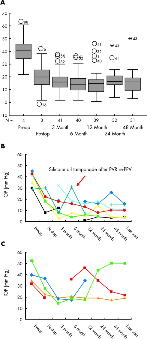
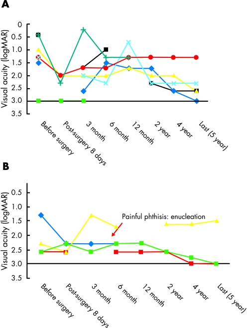
Methods: This was a consecutive interventional case series. In 44 consecutive eyes (39 patients, 22 men and 17 women) retinectomy was performed to lower the intraocular pressure (IOP) in patients with uncontrolled IOP (>35 mm Hg for more than 4 months) despite conventional filtering surgery and drug treatment. Pars plana vitrectomy was performed and the peripheral retina was surgically excised to various degrees. The procedure was concluded by an intraocular gas tamponade of 20% C(3)F(8). Included were patients with neovascular glaucoma (12 eyes), infantile and juvenile glaucoma (three eyes), secondary glaucoma due to aphakia (13 eyes), severe ocular trauma (seven eyes), uveitis (seven eyes), and glaucoma in Ehlers-Danlos syndrome (two).
Results: All patients underwent successful surgical retinectomy. All patients were followed for 5 years. Mean postoperative IOP after 4 years was 15.7 (SD 9.4) mm Hg, representing a decrease of IOP by 61% compared to the preoperative level (41.2 (9.4) mm Hg). In 52.3% of eyes long term regulation of IOP could be achieved without complications. Retinectomy was least effective in neovascular glaucoma because of central retinal vein occlusion (CRVO). Eyes with glaucoma secondary to uveitis showed a tendency towards low IOP levels with subsequent phthisis bulbi. The initial visual acuity of all patients was lower than 20/50 (mean 1.8 (0.8) logMAR) in the treated eye. Final visual acuity was 2.3 (0.6) logMAR. 21 out of 44 cases developed retinal complications (retinal detachment or proliferative vitreoretinopathy (PVR)) after surgery, requiring silicone tamponade in 11 eyes (52%) either for persistent low IOP or for PVR. Nine eyes developed phthisis, seven of which were enucleated during the follow up.
Conclusions: Long term results after retinectomy demonstrate its efficacy in otherwise intractable glaucoma. Efficacy and safety of retinectomy are dependent on the underlying disease.
Figures
Comment in
-
Retinectomy for intractable glaucoma.Br J Ophthalmol. 2004 Oct;88(10):1352-3. doi: 10.1136/bjo.2004.042820. Br J Ophthalmol. 2004. PMID: 15377568 Free PMC article. No abstract available.
Similar articles
-
Combined pars plana vitrectomy and pars plana Baerveldt tube placement in eyes with neovascular glaucoma.Retina. 2015 Jan;35(1):17-28. doi: 10.1097/IAE.0000000000000235. Retina. 2015. PMID: 25046391
-
Pars plana vitrectomy, endolaser coagulation of the retina and the ciliary body combined with silicone oil endotamponade in the treatment of uncontrolled neovascular glaucoma.Graefes Arch Clin Exp Ophthalmol. 1999 Dec;237(12):969-75. doi: 10.1007/s004170050332. Graefes Arch Clin Exp Ophthalmol. 1999. PMID: 10654165
-
Combined pars plana vitrectomy and glaucoma drainage implant placement for refractory glaucoma.Am J Ophthalmol. 2000 Mar;129(3):334-41. doi: 10.1016/s0002-9394(99)00363-3. Am J Ophthalmol. 2000. PMID: 10704549
-
Predicting visual outcome following retinectomy for retinal detachment.Br J Ophthalmol. 2008 Jul;92(7):954-8. doi: 10.1136/bjo.2007.131540. Epub 2008 Jun 12. Br J Ophthalmol. 2008. PMID: 18556423 Review.
-
Pharmacologic ciliary body ablation for chronic glaucoma in dogs: A retrospective review of 108 eyes from 2013 to 2018.Vet Ophthalmol. 2021 Mar;24 Suppl 1:125-130. doi: 10.1111/vop.12816. Epub 2020 Aug 28. Vet Ophthalmol. 2021. PMID: 32857917 Review.
Cited by
-
Strategies to influence PVR development.Graefes Arch Clin Exp Ophthalmol. 2004 Aug;242(8):699-703. doi: 10.1007/s00417-004-0978-8. Epub 2004 Aug 10. Graefes Arch Clin Exp Ophthalmol. 2004. PMID: 15309556 Review.
-
[Boston keratoprosthesis: 73 eyes from Germany : An overview of experiences from two centers].Ophthalmologe. 2018 Sep;115(9):744-753. doi: 10.1007/s00347-017-0581-0. Ophthalmologe. 2018. PMID: 29043440 German.
-
Radial Retinotomies with Endodiathermy for Severe Proliferative Vitreoretinopathy: Short-Term Results.J Ophthalmol. 2016;2016:2594574. doi: 10.1155/2016/2594574. Epub 2016 Feb 28. J Ophthalmol. 2016. PMID: 27022477 Free PMC article.
-
Anatomic and functional outcomes of retinectomy for the management of complicated retinal detachment with proliferative vitreoretinopathy.Ther Clin Risk Manag. 2015 Oct 3;11:1515-21. doi: 10.2147/TCRM.S89467. eCollection 2015. Ther Clin Risk Manag. 2015. PMID: 26491338 Free PMC article.
-
Retinectomy for intractable glaucoma.Br J Ophthalmol. 2004 Oct;88(10):1352-3. doi: 10.1136/bjo.2004.042820. Br J Ophthalmol. 2004. PMID: 15377568 Free PMC article. No abstract available.
References
-
- Cordeiro MF, Chang L, Lim KS, et al. Modulating conjunctival wound healing. Eye 2000;14:536–47. - PubMed
-
- Ayyala RS, Bellows AR, Thomas JV, et al. Bleb infections: clinically different courses of “blebitis” and endophthalmitis. Ophthalmic Surg Lasers 1997;28:452–60. - PubMed
-
- Brown RH, LH Y, Walker SD, et al. Treatment of bleb infection after glaucoma surgery. Arch Ophthalmol 1994;112:57–61. - PubMed
-
- Myers JS, Yang CB, Herndon LW, et al. Excisional bleb revision to correct overfiltration or leakage. J Glaucoma 2000;9:169–73. - PubMed
Publication types
MeSH terms
Substances
LinkOut - more resources
Full Text Sources
Medical
Research Materials
Miscellaneous