. 2003 Sep 1;171(5):2637-43.
doi: 10.4049/jimmunol.171.5.2637.

M-CSF induces vascular endothelial growth factor production and angiogenic activity from human monocytes

Affiliations

M-CSF induces vascular endothelial growth factor production and angiogenic activity from human monocytes

Tim D Eubank et al. J Immunol. .

Abstract

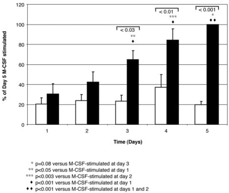
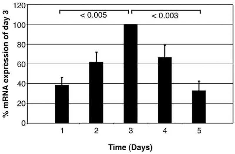
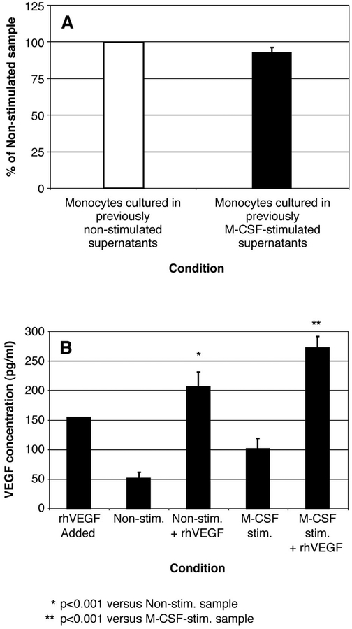
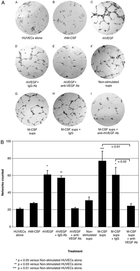
The impact of the immune response in malignancy is poorly understood. While immune cells can destroy transformed cells, the targeting and accumulation of monocytes and macrophages at tumor sites may promote tumor metastases. The growth factor M-CSF is important in promoting monocyte survival. Since M-CSF(-/-) mice are protected against tumor metastases, we hypothesized that M-CSF induced monocytes to produce angiogenic factors that facilitate metastases. In this study we demonstrate that recombinant human M-CSF induces freshly isolated normal human monocytes to produce and release the growth factor vascular endothelial growth factor (VEGF) in a dose-dependent manner, which peaked at 5 days in culture. VEGF released by these monocytes is biologically active, as cell-free supernatants from these M-CSF-stimulated monocytes induced tube formation in HUVEC. Network formation by these HUVECs after treatment with supernatants from monocytes stimulated with M-CSF were inhibited by anti-VEGF, but not by the isogenic control, Abs. Collectively, these data support an important role for M-CSF and monocytes in VEGF production and angiogenesis.


Figures

FIGURE 1.
FIGURE 1.
Kinetic analysis of VEGF production by M-CSF-stimulated monocytes. VEGF detected in supernatants over 5 days. Monocytes (5 × 10⁶/condition) were left unstimulated (☐) or were stimulated with M-CSF (100 ng/ml; ■) for the indicated time points, and VEGF in the cell-free supernatants was assayed by ELISA (picograms per milliliter). M-CSF induced more VEGF production from monocytes at 5 days than that in unstimulated, time-matched cells (*, p = 0.08 vs VEGF released from M-CSF-stimulated monocytes on day 3;**, p < 0.05 vs VEGF released from M-CSF-stimulated monocytes on day 1; ***, p < 0.003 vs VEGF released from M-CSF-stimulated monocytes on day 2; ♦, p < 0.001 vs VEGF released from M-CSF-stimulated monocytes on day 1; ♦ ♦, p < 0.001 vs VEGF released from M-CSF-stimulated monocytes on days 1 and 2). Data represent the mean ± SEM from six independent experiments.
FIGURE 2.
FIGURE 2.
M-CSF induces the expression of VEGF mRNA in human monocytes. Total RNA was isolated from monocytes (5 × 10⁶/condition) stimulated by M-CSF (100 ng/ml) for 1, 2, 3, 4, and 5 days (■) as detected by real-time PCR. M-CSF induced more VEGF mRNA at each time point and peaked on day 3 as assessed by ANOVA with post hoc testing vs unstimulated control samples at the same time (p < 0.05 for VEGF mRNA level vs unstimulated monocytes on day 3; p < 0.005 for day 3 VEGF mRNA compared with M-CSF-stimulated monocytes on day 1; p < 0.003 for day 3 VEGF mRNA compared with M-CSF-stimulated monocytes on day 5). Data represent the mean ± SEM from six independent studies.
FIGURE 3.
FIGURE 3.
There is no evidence of paracrine factors inducing VEGF in M-CSF-stimulated monocyte supernatants. A, Monocytes (5 × 10⁶/condition) were either left unstimulated (☐) or were stimulated with M-CSF (100 ng/ml; ■) for 72 h. The cell-free supernatants of these monocytes were subsequently immunodepleted of VEGF and M-CSF using specific neutralizing Abs, and the Abs were then removed by incubation with protein G agarose. The resulting supernatants were directly added to freshly isolated monocytes for an additional 72 h. Afterward, the supernatants from these monocytes were assayed for VEGF by ELISA. There was no significant difference in VEGF levels produced by the two samples after depletion (p = 0.405). Data represent the mean ± SEM calculated from four independent studies. B, Recombinant human VEGF (150 pg/ml) was supplemented back into the monocyte supernatants that were either left unstimulated (Non-stim.+ rhVEGF) or were stimulated with M-CSF (100 ng/ml; M-CSF-stim.+ rhVEGF) from A that were previously immunodepleted of VEGF and M-CSF using either anti-VEGF or anti-M-CSF neutralizing Abs (*, p < 0.001 for samples containing recombinant human VEGF added to depleted, unstimulated supernatants vs depleted, unstimulated supernatants alone; **, p < 0.001 for samples containing rhVEGF added to depleted, M-CSF-stimulated supernatants vs depleted, M-CSF-stimulated supernatants alone). These data represent the mean ± SEM calculated from four independent studies.
FIGURE 4.
FIGURE 4.
VEGF within the supernatants of M-CSF-stimulated monocytes induces network tube formation by HUVECs. A, HUVEC (1.5 × 10⁵) were grown on Matrigel as follows: A) HUVECs with RPMI medium (1 ml) alone for 20 h (HUVECs alone); B) HUVECs plus rhMCSF (100 ng/ml); C) HUVECs plus rhVEGF (10 ng/ml); D) HUVECs, rhVEGF (10 ng/ml), and isogenic IgG Ab (0.6 μg/ml; rhVEGF + IgG Ab); E) HUVECs, rhVEGF (10 ng/ml), and anti-VEGF neutralizing IgG Ab (0.6 μg/ml; rhVEGF + anti-VEGF Ab); F) HUVECs and 1 ml of supernatants from monocytes left unstimulated for 72 h (Non-stimulated sups); G) HUVECs and supernatants from 72-h M-CSF (100 ng/ml)-stimulated monocytes (1 ml; M-CSF sups); H) HUVECs, M-CSF-stimulated supernatants (1 ml), and isogenic IgG Ab (1 μg/ml; M-CSF sups + IgG); and I) HUVECs, M-CSF-stimulated supernatants (1 ml), and anti-VEGF neutralizing IgG Ab (0.6 μg/ml; M-CSF sups + anti-VEGF Ab). Pictures were taken after 20-h incubation. B, Networks of tube formation from HUVECs (1 × 10⁵/condition) that were stimulated as described above were counted in a blinded manner by adding the sum of three different fields in the well for each condition and counting completely enclosed networks. Error bars represent the mean ± SEM calculated from three independent studies. Recombinant human VEGF with or without isogenic IgG control induced endothelial tube formation (*, p < 0.05 vs unstimulated HUVECs). Similarly, supernatants from monocytes stimulated with M-CSF induced endothelial cells to form tubes (***, p < 0.01 vs unstimulated HUVECs) that were reduced by anti-VEGF, but not by isogenic control Abs (p < 0.02).

Similar articles

Cited by

References

    1. Dvorak HF, Sioussat TM, Brown LF, Berse B, Nagy JA, Sotrel A, Manseau EJ, Van De WL, and Senger DR. 1991. Distribution of vascular permeability factor (vascular endothelial growth factor) in tumors: concentration in tumor blood vessels. J. Exp. Med 174:1275. - PMC - PubMed
    1. Dvorak HF 2000. VPF/VEGF and the angiogenic response. Semin. Perinatol 24:75. - PubMed
    1. Hamada K, Oike Y, Takakura N, Ito Y, Jussila L, Dumont DJ, Alitalo K, and Suda T. 2000. VEGF-C signaling pathways through VEGFR-2 and VEGFR-3 in vasculoangiogenesis and hematopoiesis. Blood 96:3793. - PubMed
    1. Romano DP, Mangoni A, Zambruno G, Spinetti G, Melillo G, Napolitano M, and Capogrossi MC. 2002. Adenovirus-mediated VEGF(165) gene transfer enhances wound healing by promoting angiogenesis in CD1 diabetic mice. Gene Ther. 9:1271. - PubMed
    1. Senger DR, Van De WL, Brown LF, Nagy JA, Yeo KT, Yeo TK, Berse B, Jackman RW, Dvorak AM, and Dvorak HF. 1993. Vascular permeability factor (VPF, VEGF) in tumor biology. Cancer Metastasis Rev. 12:303. - PubMed

Publication types

MeSH terms